CT stands for computed tomography
Tomo- Means section
Graphy-means presentation
It’s a diagnostic device. It takes an image by applying an X-ray beam to the patient.
The way it works is by creating an x-ray beam by two detectors .gas filed detectors or
scintillation detectors.

CT is a neuroradiological investigation. It gives you a picture of your body. it shows a picture of soft tissue and the structure of the body. It can be used for many types of tests such as: It is used to test the chest, pelvis, and abdomen with an accurate image of all types of tissue.
Used to check the pain and breathing difficulties. Test some injuries like trauma Skeletal structure problems such as spinal treatment, hands and feet injuries. As it can show the small bones and the surrounding tissues.
Used also to detect any type of cancer such as lymphoma, and body parts cancers. It provides the presence of cancer, size, defined the precise place, and if there is any involvement with other body parts.
What is Computed Tomography?
Computed tomography (CT) is an imaging procedure that uses special X-ray equipment to create detailed images or scans of areas inside the body. It is also sometimes called computerized tomography or computerized axial tomography (CAT).
Each picture created during this process shows organs, bones, and other tissues in a thin “slice” of the body while CT is performed. The whole range of images produced in the whistle is like a loaf of sliced bread. You can see each piece individually, or you can see the whole loaf.
Some CT scan machines take continuous pictures in a helical fashion instead of taking a series of pictures of individual pieces of the body. Helical CT or spiral CT has several advantages over older CT techniques, it is faster, produces better quality 3-D images of areas inside the body, and better detects small abnormalities. Can put
CT is widely used in cancer as well as to help diagnose diseases and conditions of the circulatory (blood) system, such as coronary artery disease (atherosclerosis), blood vessel aneurysms, and blood clots. goes. spinal condition; kidney and bladder stones; boils; Inflammatory diseases, such as ulcerative colitis and sinusitis; and injuries to the head, skeletal system, and internal organs.
CT imaging is also used to detect deposits in adult patients with abnormal brain function or cognitive impairment who are being evaluated for Alzheimer’s disease and other causes of cognitive decline.
How does the CT Scan work?
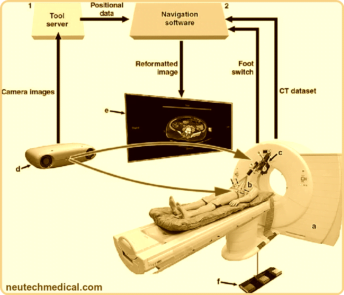
A CT-scan uses a motorized X-ray source that rotates around the circular opening of a doughnut-shaped structure called a gantry. During the CT scan the patient lies down which is slowly passed through the gantry while the X-ray tube moves around the patient. Due to which narrow rays of X-rays come out through the body. Instead of film, CT scans use special digital X-ray detectors, which are located directly opposite the X-ray source. As the X-rays leave the patient, they are detected by detectors and transmitted to a computer.
In this, each time the X-ray source completes one complete rotation, the CT computer uses sophisticated mathematical techniques to create a two-dimensional, three-dimensional image slice of the patient. The thickness of the tissue represented in each image slice can vary depending on the CT machine used, but typically ranges from 1–10 millimeters.
When a complete slice is completed, the image is stored and the motorized bed is moved forward sequentially in the gantry. Then the X-ray scanning process is repeated to produce another image slice. This process continues until the desired number of slices is collected.
These are stacked together by the computer to generate a three-dimensional image of the patient that shows the skeleton, organs and tissues as well as any abnormalities the doctor is trying to identify. This method has several advantages, including the ability to rotate the 3D image in space or view slices sequentially, making it easier to locate the exact location of the problem.
When is a CT scan needed?
CT scans are used for study by identifying disease or injury in different areas of the body. For example, CT scans of the heart are used to detect possible tumors or lesions within the abdomen, CT scans of the heart when various types of heart disease or abnormalities are suspected, including injuries, tumors, clots caused by stroke, bleeding, and others.
The position is also used for the image of the head to be detected. It can image the lungs to reveal the presence of tumors, pulmonary embolism (blood clots), excess fluid, and other conditions such as emphysema or pneumonia. A CT scan is particularly useful when imaging complex bone fractures, severely destroyed joints, or bone tumors because it usually produces more detail than a traditional X-ray.
What is the process of CT scan?
Before the scan, the patient is prohibited from eating food and asked to drink a lot of water for a specific period of time.
The day the CT scan is to be done
In most places the patient will usually have to take off their clothes, and wear a gown that the health center will provide, as well as avoid wearing jewelry.
If the hospital does not provide a gown, the patient should wear loose-fitting clothing free of metal buttons and zippers.
Some patients may need to drink contrast dye, or the dye may be given as an enema or injected. This improves the picture of blood vessels or tissues.
Any patient who is allergic to contrast material should inform the doctor in advance. Some medicines can reduce an allergic reaction to the contrast material.
Since metal interferes with the functioning of the CT scanner, the patient is required to remove all jewelry and metal fastenings.
during the scan
The patient is required to lie on a motorized table, which slides into a donut-shaped CT scanner machine.
In most cases, the patient lies on his back, facing upward. But, sometimes, they may need to lie face down or lie on their side.
After an X-ray picture, the couch will move slightly, and then the machine will take a second image, and so on, requiring the patient to lie still for best results.
During the scan, everyone except the patient will leave the room. An intercom would enable two-way communication between the radiographer and the patient.
If the patient is a child, a parent or adult may be allowed to stand or sit nearby, but they will be required to wear a lead apron to prevent radiation exposure.
Working Principle of CT Scan
It relies on target density variation that affect affects the attenuation of high-frequency waves. CT intern works onto the basic principle of X-rays which Photo-Electric Effect.
the picture of the patient is viewed thru X-ray imaging from varied angles & are then the detailed structures are mathematically reconstructed & the formed image is displayed on a video monitor.
CT Scan Vs MRI
C T works with Ionizing Radiation Effect Where M R I works with the Non-Ionizing Radiation effect. IN C T we Can View Bone Soft Tissue and a Few Nerves.
Whereas MRI we can View Bone Soft tissue and Nerves Clearly. MRI and CT both are methods of Imaging Organs of the Body, Basic Difference between both of them is that MRI will give you a clear view of the Soft Tissue of the Body organs, And CT is more preferably good for the Bone and Hard tissue.
Besides Basic Principle of Working is also different as MRI is dealing with Electromagnetic Field and CTs works on X-rays.
What I encounter is that Brain CT will not give you the clarity of All the Soft tissue of the brain in detail, Although Adjusting the image does help identify those to some extent. I don’t know, Maybe Grayscale Images of Body organs in DICOM Format.

CT & MRI both are very vital scans for Neurosurgeries relating to the Brain and Spine. Resolution & Details matter a lot, particularly in this Domain, Particularly if a surgeon has to make use of the Navigation System, He or She would need a scan with the Standard Protocols of the Navigation System.
CT and MRI are both radiographic machines but the MRI is more complicated than CT and it uses magnetic fields. The soft tissue details can be evaluated better in MRI than CT scanners.
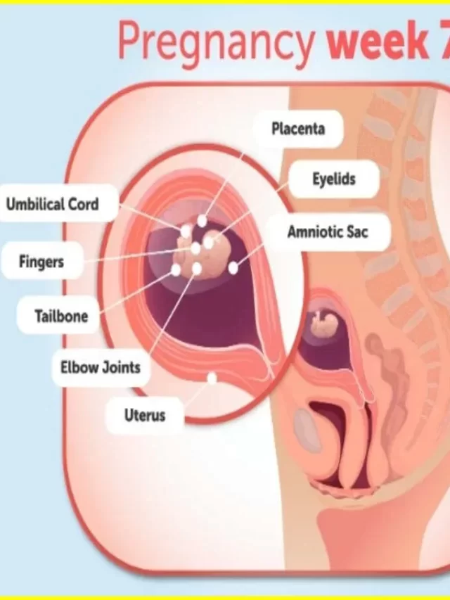
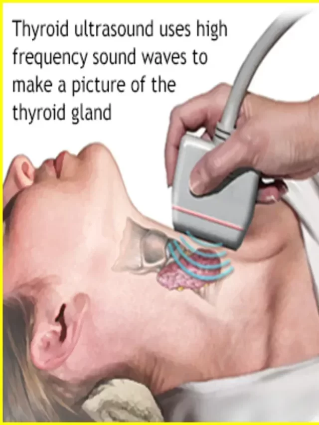
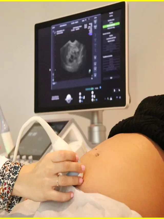
In pregnancy, other devices that do not include radiation such as MRI and ultrasound are preferred. The images provided by the CT Scanner had a three-dimensional reconstruction.
CT scans are used to help diagnose tumors, check for internal bleeding, or check for other internal injuries or damage. CT may also be used for tissue or fluid biopsy. The CT scan test is generally painless, quick and easy. Multidetector CT reduces the time taken for the patient to lie down.
Usually, an hour’s planning is required for a CT scan. Most of the time is spent in preparation. The scan takes 10 to 30 minutes or less. Usually, you can resume your activities when a healthcare provider says it is safe to do so.
Note:
Ct scan have five generation antel now
It deference by the number of slice on detector.
What are the risks of CT scan?
A CT scan can usually diagnose life-threatening conditions such as bleeding, blood clots, or cancer. Early diagnosis of these conditions can be potentially life-saving. Whereas, CT scans use X-rays, and all X-rays produce ionizing radiation. Ionizing radiation has the potential to cause biological effects in living tissues. It is a risk that increases with the number of risks added to a person’s life. However, the risk of developing cancer from X-ray radiation exposure is generally small.
A CT scan in a pregnant woman poses no known risk to the baby if the area of the body is not the abdomen or pelvis. In general, if imaging of the abdomen and pelvis is needed, doctors prefer to use exams that do not use radiation, such as an MRI or ultrasound. However, if none of them provide the required answers, or there is an emergency or other time constraints, CT may be an acceptable alternative imaging option.
In some patients, contrast agents can cause allergic reactions, or in rare cases, temporary renal failure. IV contrast agents should not be administered to patients with abnormal kidney function as they may lead to further impairment of kidney function, which may sometimes be permanent.
Children are more sensitive to ionizing radiation and have a longer life expectancy, so they have a higher relative risk of getting cancer from this type of radiation than adults. Parents may want to ask the technologist or doctor if their machine settings have been adjusted for children.
