Digital Radiograph
There are two types of digital imaging systems used in radiography computed radiography (CR) and direct radiography (DR). Digital Radiography (DR) is an advanced form of X-ray inspection that quickly produces a digital radiographic image on a computer.
This technology uses X-ray-sensitive plates to capture data during object testing, which is immediately transferred to a computer without the use of an intermediate cassette.
Benefit
1. same as digital
2. Requires follicles for the disease process
3. It reduces X-rayprint time
4. X-ray safety indicators have been improved, improving digital safety and security
basic principle
beer-lambert law When a monochromatic light source passes through a medium, the attenuation of light is proportional to the concentration of the substance in the solution.
Operating Principle
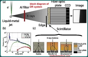
When X-ray radiation from an X-ray tube passes through the body, it falls on the X-ray detector. The detector converts the X-rays into an electrical signal and is then digitized by an analog to digital converter.
It is based on the principle that radiation is absorbed and scattered as it passes through an object.

1. A Digital Imagine
2. A digital image unit
a. An image management system
b. Image and data storage devices Interface for patient information system
c. communication network
d. A display device with controls operated by the viewer
Digital Receptor

Imaging Processing
It is also possible to use digital processing to increase the visibility of details in some radiographs. The various processing methods are explored in more detail in another module.
Digital Image Storage
Digital radiographs, and other digital medical images, are stored as digital data.
communication network
Another advantage of digital images is the ability to transfer very quickly from one place to another.
This may happen: anywhere in the world (via the Internet) for storage and display devices and other locations (teleradiology) within the imaging facility
communication network
Another advantage of digital images is the ability to transfer them very quickly from one place to another.
Digital image display and display control
Compared to radiographs recorded and displayed on film, ie softcopy, “softcopy” displays have advantages.
Basic Components of Radiography
a. X-ray tube
b. X-ray detector
c. collimator
d. HV generator
f. filter
e. console computer
x-ray tube
1. Includes cathode, anode, and casing
2. Cathode filament supply 20-160 kV. is done with
3. X-rays are generated mainly by two methods electron ejection and electron deceleration
4. Electron ejection (characteristic X-ray radiation): The fastest moving electron hits the electrons in the innermost shell, so electron vacancy is created, this vacancy is captured by electrons from the other shell emitting X-ray radiation for.
5. Electron deceleration (Bremsstralung radiation): When rapidly moving electrons approach the anode, the anode atoms appear as positive nuclei and negative electrons, thus shifting any regions that emit X-ray radiation .
6. Heat Curve: Time taken to achieve particular MA and KV
7. Cooling curve: time taken to cool the X-ray tube
8. Energy on the X-ray tube = kV mA exposure time
9. The number of electrons in the X-ray depends on the current, voltage, which improves the intensity of the X-ray beam.
HV generator
1. Used to supply X-ray tubes
2. voltage range It is mainly single phase or three phases
3. Supply specification is mainly represented by Peak kilo voltage (kVp)
4. HV: The transformer increases the input voltage.
5. Rectifier: It converts high voltage AC supply into DC supply
6. Chopper: Boosts frequencies up to 200KHZ
7. Higher frequency supply improves the maximum for exposure (KVP), provides better image sharpness and extends the life of the head tube.
Image receptor
1. It absorbs attenuated X-ray radiation in the form of an image
2. Contains Image Receptor Permanent Cassette
3. It consists of a matrix array of semiconductor (thin film transistor) devices that are in the normally closed state.
4. It switches to ON position during exposure
5. Signal from each semiconductor amplified by the pre-amplifier
6. Amplified signal given to ADC converter which converts analog signal into digital signal and then given to digital signal processor
Mainly it includes two types of Digital Radiography
1. Direct Receptor:
Composed of amorphous selenium (A-Sc) It directly converts the input X-rays into electrical signals, then analog to digital converter, this digital signal is processed by the digital processor.
2. Indirect receivers:
This input converts the X-rays first into light signals then it is passed to the photo detector, it converts the light into electrical signals. Then the analog to digital converter converts the electric signal into a digital signal and is processed by a digital processor.
collimator and grid
1. The X-ray is present between the tube and the patient.
2. Patient is exposed to collimator light prior to X-ray exposure
3. It provides proper positioning of the X-ray tube
4. Grids are lead strips inserted between the patient and the film cassette
5. It is used to reduce the contrast due to scattered radiation
This results in lower costs due to less radiation, elimination of chemical processors, processor maintenance, and filing and mailing. Less space required No dark rooms are required, and the need to dedicate space for analog images cabinets is eliminated.
Digital X-ray imaging has many advantages that benefit the clinician in less time, less radiation, the ability to take multiple exposures without changing the sensor, the storage and maintenance of images, and the electronic transmission of images.
disadvantages of Digital Radiography
The main disadvantages of direct digital radiography are the thickness and stiffness of the digital detector, temperature maintenance, infection control, hardware and software maintenance, and the high initial cost of the system.
