

Best 4d, 3d Ultrasound Pictures
Ultrasound imaging has been around for decades, but recent advances in technology have made it possible to produce 3D and even 4D images of unborn babies. These new ultrasound pictures offer a much more realistic view of the fetus than the traditional 2D images, and they have become increasingly popular in recent years. While 3D and 4D ultrasound pictures are not yet available everywhere, they are becoming more common as more hospitals and clinics invest in the necessary equipment.
If you are pregnant and considering having a 3D or 4D ultrasound picture taken, there are a few things you should know first. In this blog post, we will explore the different types of ultrasound pictures available, as well as the benefits and risks of having one taken. We will also provide some tips on how to choose a facility that offers this type of imaging.
3d Ultrasound Pictures
When you are pregnant, you will likely have multiple ultrasounds. The most common type is the abdominal ultrasound, which uses a transducer on your abdomen to create images of your baby. However, you may also have a vaginal ultrasound, in which case a small transducer is inserted into your vagina to get a better view of your baby.
Whether you are having an abdominal or vaginal ultrasound, the process is generally the same. You will lie on your back on an exam table and the technologist will apply gel to your skin. They will then move the transducer over your stomach or vagina, depending on the type of ultrasound you are having.


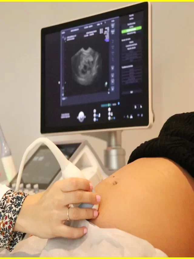


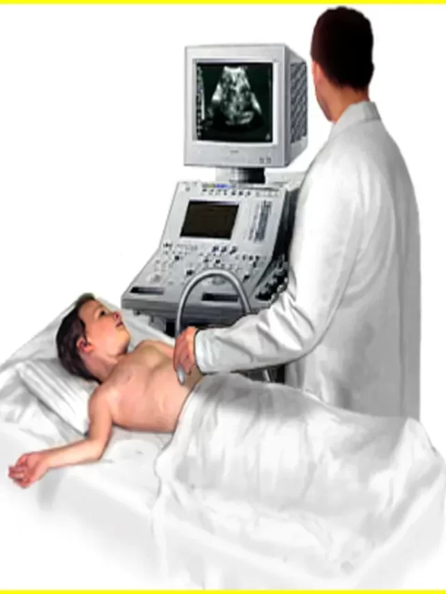
The transducer emits sound waves that create echoes. These echoes are converted into electrical signals that are then turned into images of your baby on a monitor. The technologist will take several pictures of your baby from different angles.
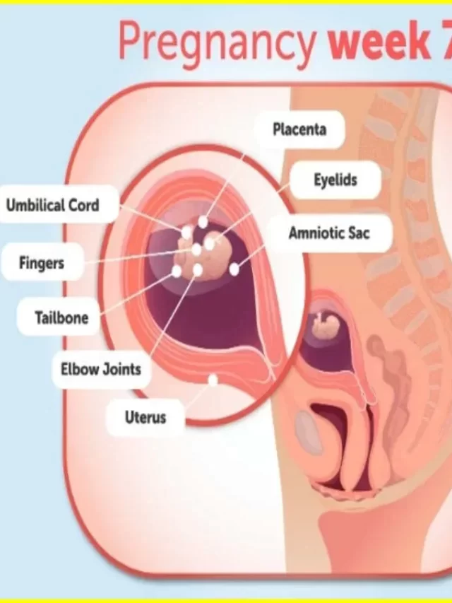
You should be able to see your baby’s heartbeat during a d ultrasound, which is usually done between weeks 14 and 20 of pregnancy. You may also be able to see other features, such as your baby’s face, limbs, and kidneys. By week 20, most babies measure about 8 inches long from head to bottom (crown-rump length). 3D ultrasound pictures
3d Ultrasound Pictures 20 Weeks
If you are pregnant and 20 weeks along, you may be wondering what your d ultrasound pictures look like. Here are some examples of d ultrasound pictures at 20 weeks.
Your baby at 20 weeks is about the size of a large cantaloupe melon. You may be able to see your baby sucking his or her thumb on the ultrasound. You may also be able to see your baby’s heartbeat.
Your baby’s organs are continuing to develop and grow. The liver and spleen begin to produce red blood cells. The kidneys begin to produce urine.
At 20 weeks, your baby’s gender can usually be determined on the ultrasound.
Here are some examples of d ultrasound pictures at 20 weeks:
Image 1:
Caption: This image shows a side view of the fetus at 20 weeks gestation. The umbilical cord is visible running from the fetus to the placenta in the lower part of the image.
Image 2:
Caption: This image is a 3D rendering of a fetus at 20 weeks gestation. The skull, spine, arms, and legs are visible, as well as the umbilical cord running to the placenta in the lower part of the image.
Image 3:
Caption: This image shows a close up view of the face of a fetus at 20 weeks gestation. You can see clearly defined features such as the eyes,
32 Week 3d Ultrasound Pictures Before and After
3d ultrasound pictures can be taken before and after the baby is born. They are a great way to see how your baby is developing and to get a glimpse of what they will look like when they are born. 3d ultrasounds can also be used to monitor the health of your baby and to check for any potential problems.
16 Weeks 3d Ultrasound Pictures
At your 3-D ultrasound, you will be able to see your baby’s facial features, including their nose, mouth, and eyes. You may even be able to see their tiny toes and fingers! These pictures are truly amazing and something you will cherish for years to come.
Article About:- Health & fitness
Article About:- Medical Technology
Article About:- IR News
Article About:-Amazon Product Review

23 Weeks 3d Ultrasound Pictures
This is an ultrasound image of a 3-week-old fetus. The gestational sac and yolk sac are clearly visible, as is the fetal pole. This image was taken during a transvaginal ultrasound.
A 3D ultrasound picture of a 3-week-old fetus will show the same structures as a 2D ultrasound, but in three dimensions. This allows for a better view of the baby and can provide more information about the baby’s development.
30 Week 3d Ultrasound Pictures
If you are wondering what a 3D ultrasound looks like, here are some pictures from different weeks of pregnancy. The first picture is from week 3 and the other two are from weeks 6 and 9.
As you can see, the 3D ultrasound pictures show more detail than the 2D ultrasound pictures. You can see the baby’s head, arms, and legs more clearly. The 3D pictures also show the baby’s movements better.
2022 Newest Ultrasound Machine for Pregnancy Portable ecografo portatil Scanner Handheld Doppler for Small Baby Dog sonogram ultrasounds veterinario Veterinary Portable Ultrasound for Bladder Dogs


