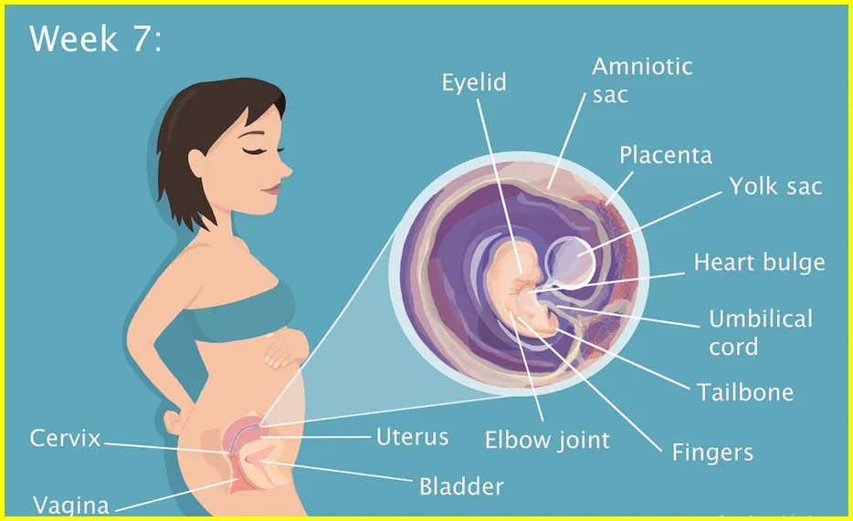
Ultrasound Week 7 What to expect
If you’re pregnant and wondering what to expect during your 7 week ultrasound, you’ve come to the right place. Here’s an overview of what you can expect during this important milestone in your pregnancy.
Ultrasound Week 7
Ultrasound week 7, you will be able to see your baby’s heart and all four chambers. The size of the ventricles and atria should be the same. The valve between the chambers must be open and working properly. You can also see the beginnings of the coronary arteries.
7 Week Ultrasound Twins
The ultrasound is a special test that uses sound waves to create a picture of your baby (or babies). It’s also called a sonogram.
First trimester to confirm your due date, check multiple pregnancies, and assess the risk of certain birth defects. This ultrasound is usually done between 10 weeks and 13 weeks + 6 days of gestation.
Second trimester to evaluate fetal growth and development, and to look for any physical problems. This ultrasound is usually done between 18 weeks and 20 weeks + 6 days of gestation.
Third Trimester to check your baby’s position in the womb (birthing canal), assess the location of the placenta, listen to the heart rate, and measure the fluid around the baby. This ultrasound is usually done between 32 weeks and 34 weeks + 6 days of pregnancy. However, some physicians will perform another scan at 36 weeks’ gestation to re-assess the position of the fetus and estimate weight.
If you’re pregnant with twins or more, you may have additional ultrasounds to check their growth and development compared to other babies of the same gestational age. These “developmental scans” are usually done every 4 weeks from 28-32 weeks gestation until delivery.
An ultrasound uses sound waves to create an image of your baby on a screen. The amount of time this takes depends on how cooperative your little one is—and how many babies you’re carrying! Jail during exam.

What Does a 7 Week Ultrasound Look Like
When you are Ultrasound Week 7 pregnant, your baby is about the size of a kidney bean. At this stage of development, ultrasounds can provide clear information about your baby’s head, spine, arms and legs. You can also see your baby’s heartbeat on the screen.
Normal 7 Week Ultrasound
Expect to see a lot of movement during your baby’s ultrasound.
He’ll be very active, and you may even see him sucking his thumb.
All organs and major body parts should be visible, and the doctor will look for any abnormalities.
The baby’s heart will be beating rapidly – about 150 times per minute.
You can also hear its heartbeat during the ultrasound.
At this stage, the baby is about the size of a kidney bean and weighs about half an ounce.
7 Week Ultrasound HeartBeat
The ultrasound week is an important time to monitor your baby’s health. The main goals of this ultrasound are to check the baby’s heart rate and assess the overall health of the pregnancy.
During the ultrasound, the technician will place a transducer on your abdomen. This transducer emits sound waves that create an image of your baby on a screen. The technician will use this image to check the baby’s heart rate and look for any abnormalities.
It is normal for the baby’s heart rate to fluctuate during this ultrasound. However, if the heart rate continues to drop, it could be a sign of a problem. The technician will closely monitor the heart rate and provide you with information on what to do next if you have any concerns.
After the ultrasound, you will be informed about the results. If everything appears normal, you can continue with your pregnancy as planned. However, if there is any concern, you may need to follow up with additional ultrasounds or other tests.
Article About:- Health & fitness
Article About:- Medical Technology
Article About:- Sports

Baby 7 Week Ultrasound
The Ultrasound Week 7 can be one of the most exciting moments during pregnancy. This is when you can see your baby for the first time and find out the gender. An ultrasound can also help determine how far along you are in your pregnancy. Here’s what you can expect during your baby’s 7-week ultrasound.
During a baby 7 week ultrasound, the technician will apply a gel to your abdomen and use a transducer to get a clear image of your baby. They will measure your baby’s size and look at the heart, brain, spine, kidneys and liver. You can see your baby move and hear the heartbeat. The ultrasound will take about 30 minutes.
If you have any questions or concerns about the 7-week ultrasound, talk with your doctor or the ultrasound technician.
Are the twins appearing so soon?
Yes, especially if they are fraternal. This is one of the main reasons for an early ultrasound to find out how many babies are growing in your uterus.
If you have fraternal twins – meaning, two different eggs were fertilized – there will be a different gestational sac for each baby. If you’re estimating your pregnancy correctly, several sacs should be clearly visible on the transvaginal Ultrasound Week 7.
If your twins are identical – meaning, one egg was fertilized but then split in two – there will be only one gestational sac; However, more than one yolk sac, embryonic pole, and heartbeat may be visible.
Again, keep in mind that ultrasounds are not foolproof. You may not be far enough along in your pregnancy to figure all these things out.
And remember that kids love to hide, especially when they have a sibling to hide behind. Many gestational sacs are not visible until a later ultrasound.
How will my 7 week ultrasound be done?
This early baby scan is usually done across the abdomen. However, in some cases, an internal scan (transvaginal) may be needed to see all the necessary details or if your womb is tilted backwards.
We will always try to do a trans-abdominal scan first but if we need to do a transvaginal ultrasound scan we will discuss this with you. To perform this early baby scan, you will be asked to lie on the examining couch and expose your lower abdomen.
A small amount of gel will be applied to your skin. The gel will help the transducer make good contact with the skin. The ultrasound transducer will be placed on the body and moved in different directions over the area of interest to obtain the required information/ultrasound images.
There is usually no discomfort from the pressure in the form of the transducer or probe. However, if the scanning is done over an area of tenderness, you may feel pressure or slight discomfort from the transducer.

Once the ultrasound scan is complete, the clear ultrasound gel will be rinsed off your skin. Any part that is not wiped will dry quickly. Ultrasound Gel generally does not stain or discolor clothing.
This ultrasound exam is usually completed within 10-15 minutes. After an ultrasound exam, you should be able to resume your normal activities right away.
What are the benefits and risks of an early pregnancy ultrasound exam?
Benefits
- Early pregnancy, the reassurance scan is non-invasive.
- If a transvaginal scan is required then the ultrasound examination may be slightly uncomfortable but not painful.
- Ultrasound examinations are fairly inexpensive when compared to other diagnostic imaging methods.
- Ultrasound imaging is safe for the baby and the mother because there is no ionizing radiation involved.
- You can’t see the baby using conventional X-ray imaging.
- Ultrasound is the preferred imaging test for the diagnosis and monitoring of pregnancy.
- Ultrasound allows the sonographer to see the inside of the uterus in real time and provides essential information about the pregnancy.
Risk
- There are no known harmful effects on humans or babies related to pregnancy ultrasound examinations.
- Although ultrasound has been used in pregnancy for more than 40 years, there is no evidence that it is harmful to the patient, embryo or fetus, ultrasound should be performed only when medically indicated and by qualified practitioners done by.
what does a 7 week ultrasound look like
If you’re pregnant and wondering what to expect during your Ultrasound Week 7, you’ve come to the right place. Here’s an overview of what you can expect during this important milestone in your pregnancy.
what to expect at 7 week ultrasound
If you’re pregnant and wondering what to expect during your Ultrasound Week 7, you’ve come to the right place. Here’s an overview of what you can expect during this important milestone in your pregnancy.
what does 7 week ultrasound look like
Ultrasound week 7, you will be able to see your baby’s heart and all four chambers. The size of the ventricles and atria should be the same. The valve between the chambers must be open and working properly. You can also see the beginnings of the coronary arteries.
what can you see at 7 week ultrasound
When you are Ultrasound Week 7 pregnant, your baby is about the size of a kidney bean. At this stage of development, ultrasounds can provide clear information about your baby’s head, spine, arms and legs. You can also see your baby’s heartbeat on the screen.
what should a 7 week ultrasound look like
When you are Ultrasound Week 7 pregnant, your baby is about the size of a kidney bean. At this stage of development, ultrasounds can provide clear information about your baby’s head, spine, arms and legs.
Can you see anything on a 7 week ultrasound?
At the Ultrasound Week 7 scan, only a gestational sac and yolk sac may be seen. It’s still very early in the pregnancy. If there are concerns, you may be asked to return for another scan in 7 to 10 days to check on the embryo’s development.
Can you see a heartbeat at 7 week ultrasound?
A strong fetal heartbeat can be clearly seen at 7 weeks. The range can be from 100 to 180 beats per minute (bpm) . Any earlier than Ultrasound Week 7, you may not see the embryo or fetal heart beating due to the embryo being so small.
Is no heartbeat at 7 weeks normal?
No Fetal Heartbeat After Seven Weeks Gestation
If you are past seven weeks pregnant, seeing no heartbeat may be a sign of miscarriage.1 By this point a transvaginal ultrasound should be able to reliable detect a heartbeat or lack thereof. But there are many exceptions to the “heartbeat by seven weeks” rule.
Is a heartbeat at 7 weeks good?
Generally, from 6 ½ -7 weeks is the time when a heartbeat can be detected and viability can be assessed. A normal heartbeat at 6-7 weeks would be 90-110 beats per minute. The presence of an embryonic heartbeat is an assuring sign of the health of the pregnancy.
How do I know my baby is OK 7 weeks?
Brace yourself to give a variety of samples (blood, urine and cervical cells for a pap smear), get an ultrasound that will confirm baby’s doing okay in there, and get an estimated due date (yep, you might already have one, but the doctor may adjust it a bit based on what they see).
WavHello BellyBuds Baby Bump Headphones – Prenatal Belly Speakers for Women During Pregnancy, Safely Play Music, Sounds, and Voices to Your Baby in The Womb – Green


