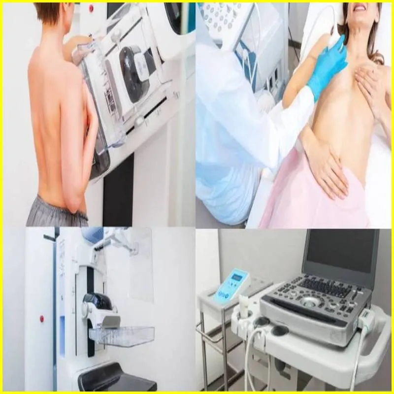
What is the Breast Ultrasound Procedure, Purpose
A breast ultrasound is a diagnostic tool used to create an image of the breasts. The procedure is sometimes called a sonogram. The test uses high-frequency sound waves to create pictures of the inside of the breast. It is generally used to check for abnormalities such as lumps, cysts or masses. A breast ultrasound is usually performed as an outpatient procedure. This takes about 30 to 60 minutes.
Breast Ultrasound
A breast ultrasound is a diagnostic test that uses sound waves to create an image of the inside of the breast. It is used to evaluate masses or lumps that cannot be seen on a mammogram. Ultrasound is also used to help guide a needle biopsy of a suspicious area.
The test is performed by a radiologist, who will apply a gel to the breast and then place a hand-held device called a transducer against the skin. The transducer emits sound waves that bounce off structures inside the breast and are converted into electrical signals. These signals are then displayed as images on a computer screen.
Breast ultrasound is considered safe and does not carry any risk. However, false-positive results are possible, meaning that an abnormal result may be found even though no cancer is present. If you have had a breast ultrasound, you should contact your doctor to discuss the results and whether additional testing is needed.
Article About:- Health & fitness
Article About:- Medical Technology
Article About: Sports

Breast Ultrasound vs Mammogram
Breast ultrasound is an imaging test that uses sound waves to create a picture of the inside of your breast. It is used to check for problems such as cysts, tumors, or other abnormalities.
A mammogram is an X-ray test of the breast. It is used to look for changes that may be cancerous or noncancerous, such as lumps, calcium deposits, and abnormal tissue growth.
Both procedures are important tools for diagnosing breast problems, but they have different strengths and weaknesses.
Breast Ultrasound vs Mammogram: Procedure
During a breast ultrasound, you lie on your back on an exam table. A gel is applied to your skin, and a handheld device called a transducer is moved to your breast. The transducer emits sound waves that bounce off structures in your breast and create echoes. These echoes are converted into images which are displayed on the monitor. The test usually takes less than 30 minutes.
Mammograms also involve lying on an exam table, but you place your breast on a flat surface called a compression plate. The plate squeezes your breast so it’s flatter and easier to image. An X-ray machine then takes pictures of your breast from different angles. The test takes 10-15 minutes per breast.
Breast Ultrasound vs Mammogram: Purpose
Normal Breast Ultrasound
A breast ultrasound is a diagnostic imaging test that uses high-frequency sound waves to create pictures of the internal structures of the breast. Ultrasound images are often used to complement mammography, which is the gold standard for breast cancer screening.
Breast ultrasounds are usually performed as outpatient procedures and take about 30 minutes to complete. The test is usually performed by a radiologist, a medical doctor who specializes in interpreting medical images.
A technologist will apply a gel to the breast and then hold a handheld device called a transducer against the skin. The transducer emits sound waves that bounce off the breast tissue and produce echoes. These echo signals are converted into electrical impulses that form an image of the breast on a computer screen.
Diagnosis and Screening. Diagnostic ultrasound is used to evaluate abnormal findings on a mammogram or physical exam, such as a lump or mass. Screening ultrasound is used to screen women who have no symptoms or signs of breast cancer but are at high risk because they have a strong family history of the disease or other risk factors, such as dense breasts.
Both types of ultrasound use the same equipment and techniques. The main difference is that diagnostic ultrasounds are more focused and take longer to perform than screening ultrasounds.
There are no known risks associated with having a breast ultrasound. However, the procedure may be uncomfortable for some people due to the pressure of the transducer.
Abnormal Breast Ultrasound
An abnormal breast ultrasound may be concerning, but does not mean you have cancer. Here’s what you need to know about this diagnostic tool.
Breast ultrasound is used to visualize the breasts and look for abnormalities. The test is carried out by a radiologist, who will take images of the breasts using a special ultrasound machine.
Ultrasound is safe and painless, and it does not use ionizing radiation (like mammograms do). This makes it a good choice for pregnant or lactating women.
Abnormal breast ultrasounds are relatively common. In fact, 1 in 4 women who get a mammogram will also need an ultrasound. Most of the time, an abnormal ultrasound is due to benign (non-cancerous) conditions such as cysts or fibroids.
However, in some cases, an abnormal breast ultrasound may be a sign of cancer. If your radiologist sees something suspicious on your ultrasound, they may recommend additional testing with a mammogram or biopsy.
It is important to remember that most breast abnormalities are not cancer. However, if you have any concerns about your results, be sure to discuss them with your doctor.

Breast Ultrasound Cost
- The cost of a breast ultrasound procedure can range from $200 to $1000 depending on the provider and location.
- Insurance companies usually cover the cost of breast ultrasounds if they are deemed medically necessary.
- Some insurance plans may require a co-pay or deductible for the procedure.
- There are several ways to reduce the cost of a breast ultrasound, including using a provider that is in network with your insurance company or negotiating a lower price with your provider.
what does breast cancer look like on ultrasound

A breast ultrasound is a diagnostic test that uses sound waves to create an image of the inside of the breast. It is used to evaluate masses or lumps that cannot be seen on a mammogram. Ultrasound is also used to help guide a needle biopsy of a suspicious area.
how accurate is ultrasound in detecting breast cancer

The test is performed by a radiologist, who will apply a gel to the breast and then place a hand-held device called a transducer against the skin. The transducer emits sound waves that bounce off structures inside the breast and are converted into electrical signals. These signals are then displayed as images on a computer screen.
What will a breast ultrasound show?

Breast ultrasound uses sound waves and their echoes to make computer pictures of the inside of the breast. It can show certain breast changes, like fluid-filled cysts, that can be harder to see on mammograms.
Can you detect breast cancer with an ultrasound?

A breast ultrasound is most often done to find out if a problem found by a mammogram or physical exam of the breast may be a cyst filled with fluid or a solid tumor. Breast ultrasound is not usually done to screen for breast cancer. This is because it may miss some early signs of cancer.
Why would you need a breast ultrasound?

If you feel a lump in your breast, or one shows up on your mammogram, your provider may recommend an ultrasound. A breast ultrasound produces detailed images of breast tissue. It can reveal if the lump is a fluid-filled cyst (usually not cancerous) or a solid mass that needs more testing.
what breast cancer looks like on ultrasound

A brest ultrasound is most often done to find out if a problem found by a mammogram or physical exam of the breast may be a cyst filled with fluid or a solid tumor. Breast ultrasound is not usually done to screen for breast cancer. This is because it may miss some early signs of cancer.
Is breast ultrasound painful?

Does a Brest Ultrasound Hurt? Breast ultrasounds should never feel painful or uncomfortable. However, you may feel slight discomfort if the transducer scans over a sensitive or tender area of your breast. Because ultrasound technology doesn’t use radiation, breast ultrasounds are even safe to use during pregnancy.
Can ultrasound detect breast cysts?

This test can help your doctor determine whether a breast lump is fluid filled or solid. A fluid-filled area usually indicates a breast cyst. A solid-appearing mass most likely is a noncancerous lump, such as a fibroadenoma, but solid lumps also could be brest cancer.
When is the best time to have breast ultrasound?

Consider scheduling your breast exam one to two weeks after your period begins. Breasts are the least tender just after menstruation, making the exam more comfortable.
SZEMENTMD Breast Ultrasound Puncture Model EMM-RA, 4 Simulated lesions Inside for Teaching and Practical Training of Breast biopsy, Minor Lesion Location, Rotational Resection of Benign lesions
[Breast Ultrasound Puncture Training] 4 Simulated lesions of varying sizes are present inside the tissue in the ultrasound-guided deep tissue mass puncture training model for practice, a unique tool for your practical training and exercises that test your associated skills.
[Excellent ultrasonic guidance performance] It adopts polymer materials equivalent to human tissue, and ultrasonic development is equilalent to human tissue density. Different gray-scale acoustic images are displayed in different tissues when ultrasound guidance is used, so as to distinguish lesions from normal tissues.
[Simulate breast biopsy] It adopts polymer materials equivalent to human tissue, and ultrasonic development is equivalent to human tissue density.
[Simulate minor lesion location] It contains 1cm diameter hypoechoic nodule tissue and 1.5cm hypoechoic malignant invasive lesion tissue, which are located in different directions and different spatial distributions of the breast.
[ Simulate benign lesion excision] Random spatial distribution of 4 lesions with different sizes.


