Where are Transmission Electron Microscopes Used?
Transmission electron microscopes (TEMs) produce images that are greatly enlarged by using an electron particle beam to view specimens. Up to a million times magnification is possible with TEMs. Consider how little a cell is to have a better understanding of how tiny that is.
Ever questioned how scientists detect little particles that are invisible to the human eye? An extremely potent device called a transmission electron microscope (TEM) holds the key to the solution. The definition of TEMs and its numerous, fascinating uses in fields like manufacturing, research, and medical will be discussed in this article.
What is the working principle of Transmission electron microscopes (TEMs)
Transmission electron microscopes, or TEMs, are strong tools for high-resolution nanoscale imaging of thin materials. TEMs operate on the basis of using electrons rather than light for imaging, which provides far greater resolution than that of conventional light microscopes.

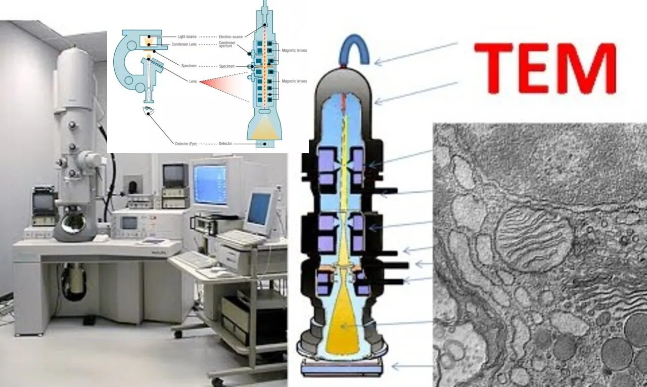
This is a condensed description of how Transmission Electron Microscopes operate:
- Electron Source:
- TEMs use an electron gun to produce a beam of electrons. This electron gun typically consists of a heated tungsten filament or a field emission cathode. The high temperature or electric field applied to these sources causes electrons to be emitted.
- Electron Lenses:
- Once emitted, the electron beam passes through a series of electromagnetic lenses. These lenses focus and shape the electron beam, similar to how glass lenses in an optical microscope focus light. Magnetic coils generate the electromagnetic fields that control the path of the electrons.
- Condenser Lens:
- The condenser lens focuses the electron beam onto the specimen. This focused beam is used to illuminate the sample.
- Specimen:
- The specimen in a TEM is an extremely thin section of the material of interest. Since electrons have very short wavelengths, they can interact with the specimen at the atomic level, providing high-resolution images.
- Transmission through the Specimen:
- The electron beam passes through the specimen, and some electrons are scattered or absorbed by the specimen’s atoms. The degree of interaction depends on the thickness and composition of the specimen.
- Objective Lens:
- The transmitted electrons then pass through an objective lens, which forms a magnified and focused image of the specimen onto a fluorescent screen or a digital camera. This image is a result of the electrons that have passed through the specimen without being scattered or absorbed.
- Magnification and Imaging:
- The final image is formed on a detector, and the magnification achieved in TEMs is much higher than that of optical microscopes, allowing for the visualization of structures at the atomic level.
- Additional Components:
- TEMs often include other components such as a vacuum system to minimize electron scattering, specimen holders for precise positioning, and various detectors for specific types of imaging and analysis.
Transmission Electron Microscopes in the Medical Field
The medical profession is one of the main applications for the transmission electron microscope (TEM). As well as aiding in illness diagnosis, TEM has been crucial in the creation of several vaccinations and therapies.
Viruses are too tiny to view under a light microscope, but TEM makes them visible. It has been crucial to our knowledge of how viruses function and to the creation of vaccinations to protect against them. For instance, the structure of the poliovirus was studied using TEM, which helped to produce the polio vaccine.
Additionally, cells from individuals with probable malignancy may be examined using TEM. Doctors can frequently determine the type of cancer and if a patient has it by examining at cells under TEM. Determining each patient’s optimal treatment approach requires consideration of this information.
TEMs are utilized for research into novel medications and treatments in addition to their function in the diagnosis and treatment of illnesses. Through the use of TEM, scientists may better understand the genesis and progression of illnesses by examining cells and tissues. Having this knowledge is crucial to creating novel, successful therapies.
Article About:- Health & fitness
Article About:- Medical Technology
Article About:-Sports

The Applications of a transmission electron microscopes
A transmission electron microscope (TEM) is an instrument that uses a beam of electrons to produce a detailed image of a sample. TEM is used in many scientific fields to study the structure and properties of materials at the atomic level.
Some of the most common applications of TEM include:
- Materials science: TEM is used to analyze novel materials’ characteristics and investigate their microscopic structure.
- Biology: TEM is used to examine the mechanics of cell division and death as well as the structure of viruses and cells.
- Nanotechnology: The research and creation of nanomaterials like carbon nanotubes and quantum dots are done by TEM.
- Metallurgy: TEM is used to examine corrosion and other metallurgical processes as well as the microscopic structure of metals and alloys.
How does the transmission electron microscopes work?
TEM is an imaging device that uses a high-energy electron beam to image the internal structure of a sample. TEM creates images of the sample which are magnified thousands of times. TEM can also be used to measure the dimensions of small features and to determine the chemical composition of samples.
Why use a transmission electron microscopes?
There are many reasons to use a TEM, but some of the most common reasons include:
- To acquire a sample’s high-resolution pictures
- the process of determining a sample’s atomic structure
- To examine a sample’s makeup
- to investigate the dynamics of the processes taking place in a sample
- to carry out comparative research using additional microscopy methods

What are the limitations of a transmission electron microscopes?
Using a transmission electron microscope (TEM) has several drawbacks. First and foremost, the apparatus must be able to produce an extremely powerful magnetic field, which might be costly and required specific equipment. Secondly, in order for electrons to flow through the sample, it needs to be finely cut, which might be challenging or impossible with some materials. Lastly, TEM can only be used to examine a sample’s surface; it is unable to reveal details about its inside.
Strengths and Limitations of the Transmission Electron Microscopes
Despite the fact that transmission electron microscopy is a very adaptable technology with a broad range of applications, it is crucial to recognize the limitations of this technique in light of its numerous advantages. Microscopists can create novel methods that overcome some of the limits of TEM and STEM, therefore increasing the technique’s usefulness, in addition to better comprehending the complicated data produced by these instruments.
Advantages
- offers the greatest and strongest magnification possible for a microscopy method.
- Adaptable imaging modes: high-angle annular dark field (STEM); phase contrast (TEM); dark/bright field.
- enables the use of selective area diffraction (SAD) to gather electron diffraction patterns, or crystallographic data, from areas of a nanoscale dimension.Facilitates
- The capacity to gather local data on structure and binding that can be connected to high-resolution pictures is known as nano-analysis.
Disadvantages
- Limited Sampling: An HR-TEM image’s normal field of view is no larger than 100 nm2.
- Interpreting Complex Images: Every TEM picture, including diffraction patterns, is a 2D projection of a 3D structure.
- particularly vulnerable to soft materials, biological samples, and light components being damaged by electron beams. This often makes gathering useful data prior to the sample being unsampled difficult.
- Beam-sensitive materials may be analyzed utilizing specialized methods and cutting-edge tools including cryo-TEM, low dose TEM, and low-kV TEM.
- The necessity of a vacuum atmosphere restricts the possibility of seeing functionalized materials under “working” settings that are close to reality.
- Materials under stimulus (heat, gas, liquid, electrical bias) can be studied using E-TEM and in situ TEM holders.
- ultra-thin specimens need challenging specimen preparation; thin samples frequently produce imaging artifacts.

FAQ
What is transmitted in an electron microscope?
A method known as transmission electron microscopy (TEM) involves passing an electron beam through an extremely thin specimen and seeing how the specimen reacts to the electron beam.
What are transmission electron microscopes used to view?
Using a transmission electron microscope, one may see thin materials (molecules, tissue slices, etc.) that allow electrons to flow through and produce a picture. There are several similarities between the traditional (compound) light microscope and the TEM.
What is difference between SEM and TEM?
The primary distinction between TEM and SEM is that TEM employs transmitted electrons—that is, electrons traveling through the sample—to form a picture, whereas SEM uses reflected or knocked-off electrons to do so.
Who invented transmission electron microscope?
The electron microscope is ascribed to German electrical engineer Ernst Ruska. The first commercially viable, mass-produced electron microscope was developed in 1931, and it was made available for purchase in 1939.
Why is a transmission electron microscope?
TEM may be used to examine semiconductor flaws, layer composition, and growth. The quality, shape, size, and density of quantum wells, wires, and dots may all be examined at high resolution. The TEM employs electrons rather than light to function according to the same basic principles as a light microscope.
What is the principle of TEM and SEM?
SEM creates images by scanning the sample, which is at the bottom of the electron column, and analyzing the electron scattering that results. The sample is positioned in the center of the TEM, where electrons flow past it before being collected.
What is the function of TEM?
Transmission electron microscopes (TEMs) produce images that are greatly enlarged by using an electron particle beam to view specimens. Up to a million times magnification is possible with TEMs.


