The Brain Mri With and Without Contrast Uses As A Diagnostic Tool
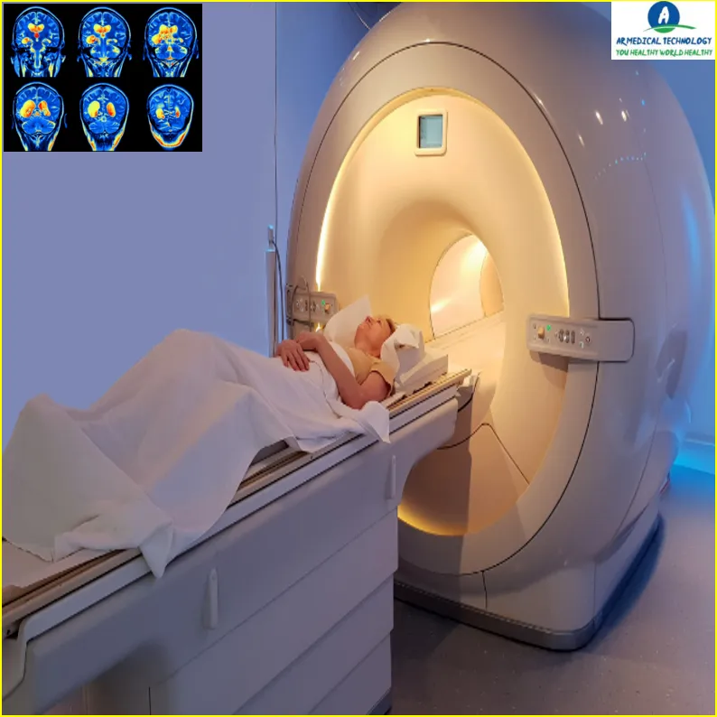
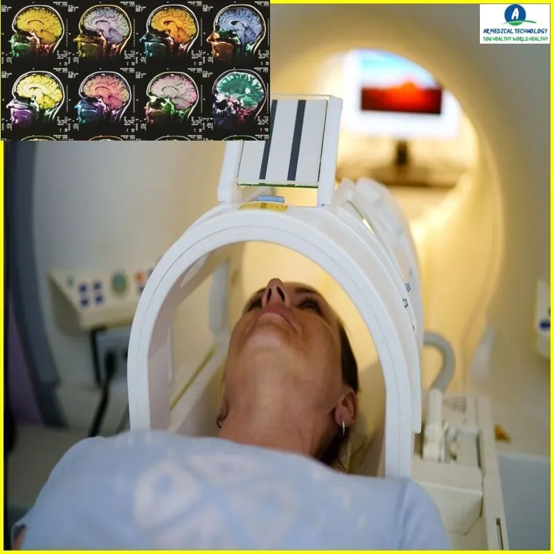
Brain MRI, also known as magnetic resonance imaging, is a diagnostic technique that creates precise pictures of your brain using radio waves and strong magnets. A number of illnesses, such as brain tumors, strokes, aneurysms, Alzheimer’s disease, and multiple sclerosis, can be diagnosed by magnetic resonance imaging (MRI). They can also be used to assess any damage resulting from an earlier collision. We’ll look at the many applications of brain MRI as a diagnostic tool in this blog article. We’ll also go over the procedure’s workings and what to anticipate when it’s over.
Brain Mri With and Without Contrast
Brain MRIs come in two varieties: contrast-enhanced and contrast-less. Contrast can help make some parts of the brain easier to see, and it is often administered via an IV. Doctors can still see a clear image of the brain in the absence of contrast, although certain aspects could be harder to discern.
CT scan.
Computed tomography (CT) scans are a kind of imaging test that employ X-rays to produce finely detailed images of the inside of the body. To inspect the brain and search for any unusual growths or alterations, a CT scan may be performed.
Article About:- Health & fitness
Article About:- Medical Technology
Article About:- Sports

EEG
An EEG, or electroencephalogram, is a test that measures the electrical activity of the brain. This test can help doctors diagnose epilepsy and other conditions that affect the electrical activity of the brain.
PET scan
A PET scan, or positron emission tomography scan, is a type of imaging test that uses special cameras and radioactive tracers to create detailed pictures of the inside of the body. A PET scan may be used to examine the brain and look for any abnormal growths or changes.
spect scan
A SPECT scan, or single photon emission computed tomography scan, is a type of imaging test that uses special cameras and radioactive tracers to create detailed pictures of the inside of the body. A SPECT scan may be used to examine the brain and look for any abnormal growths or changes.
Cpt Brain Mri With and Without Contrast
A brain MRI with and without contrast can provide a wealth of information for doctors diagnosing a variety of conditions. The scan can show things like tumors, aneurysms, strokes, and other abnormalities. It may also help to rule out certain conditions.
Contrast is usually used when something specific, such as a tumor, is suspected. By injecting a contrast agent into the bloodstream, doctors can better see how blood is flowing to different parts of the brain. This can be helpful in determining if there are any blockages or other problems.
Brain MRIs are painless and safe, and they provide a non-invasive way to view the brain in detail. They are often used in conjunction with other tests, such as an EEG or CT scan, to get a complete picture of what is happening with the patient.
Brain Mri With and Without Contrast Cpt
Brain MRI with and without contrast is a diagnostic tool that can be used to detect a variety of conditions. MRI uses magnetic fields and radio waves to create images of the inside of the body. MRI can be done with or without contrast. Contrast is a dye that is injected into the body and helps improve the quality of the image.
Brain MRI is a non-invasive procedure that does not involve any radiation. The procedure is generally well tolerated by patients and takes approximately 30 minutes to complete.
Cpt code for Brain Mri With and Without Contrast
A brain MRI (magnetic resonance imaging) is a diagnostic tool used to evaluate the structure and function of the brain. This can be done with or without contrast.
The CPT code for Brain MRI with Contrast is 70450. The CPT code for a brain MRI without contrast is 70451.

FAQ
Which is better MRI with or without contrast?
An MRI with contrast is necessary to determine whether the patient receiving therapy for a tumor is receiving it appropriately. In general, an MRI without contrast cannot be used to assess the specific tumor state. MRI contrasted pictures are more readable than contrast-free MRI images.
Is MRI with contrast safe for brain?
It’s not always a good idea to get an MRI with contrast. Gadolinium, for instance, has a difficult time passing across the blood-brain barrier (BBB), which shields the brain from dangerous chemicals.
Will an MRI without contrast show nerve damage?
One of the greatest imaging techniques for detecting soft tissue injury is an MRI; however, it is not able to detect spinal cord compression or other problems.
Can brain MRI without contrast detect tumor?
An MRI without contrast, for instance, may be used to see a big tumor; but, an MRI with contrast will be able to spot a smaller tumor and provide more accurate measurements of the tumor’s size and extent into the surrounding tissues.
What are the side effects of an MRI without contrast?
MRIs performed without contrast or anesthesia have no side effects. First, be aware that if an MRI is performed without contrast or anesthesia, there are no potential side effects or difficulties. An enormous magnet is an MRI. It uses no radiation and produces a very detailed image.
Is MRI contrast risky?
For the majority of people, MRI contrast is safe. However, before having an MRI with contrast, patients with renal illness and pregnant women should inform their physician. Generally minor, common adverse effects of contrast materials include rash, nausea, and vomiting.
Is MRI with contrast painful?
To ensure there are no surprises, we provide you with all the information you want before to the treatment. An MRI is a painless, noninvasive exam. There’s more good news: unlike CT and X-rays, radiation is not a factor.
What is the side effect of brain MRI?
The MRI scan itself doesn’t cause any negative effects. However, you could experience certain adverse effects, such as a skin rash, headache, nausea, and dizziness, if you received an injection of contrast media (dye) as part of the examination.


