scanning electron microscopes
Where are scanning electron microscopes
SEMs are one of the most often used equipment for viewing and evaluating the microscopic and nanoscopic properties of things. In this post, we’ll look at how scanning electron microscopes may help scientists get a deeper knowledge of the world around them. Learn how powerful microscopes are changing our lives in fields ranging from medical research to materials science.
What are scanning electron microscopes
SEMs are sophisticated tools that allow scientists to examine the structure of a wide range of materials in great detail. They are often employed in research laboratories to investigate the characteristics of semiconductors, metals, and other materials.
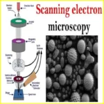
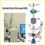
Scanning electron microscopes function by directing an electron beam onto the surface of a sample. Secondary electrons are produced when the electrons interact with the atoms in the sample. The microscope detects these secondary electrons and provides a picture of the sample’s surface.
SEMs may be used to investigate both the macroscopic and microscopic properties of a material. They are particularly excellent for analyzing surfaces at extremely high resolutions, making them an indispensable tool for a wide range of scientific inquiry.
Article About:- Health & fitness
Article About:- Medical Tech
Article About:-Sports

Uses of Scanning Electron Microscopes
Scanning electron microscopes are used in a wide range of industries and applications. They are commonly used in the semiconductor industry for quality control and failure analysis, as well as in the medical field for the study of cells and tissues.
In the semiconductor industry, scanning electron microscopes are used to examine the surface of semiconductor wafers for defects. They can also be used to analyze the structure of individual transistors and other components. In failure analysis, SEMs are used to identify the cause of defects in electronic devices.
In the medical field, scanning electron microscopes are used to study cells and tissues. They can be used to examine cell structures, such as the nucleus, and to investigate disease processes.
How does a scanning electron microscope work?
A scanning electron microscope (SEM) is a device that uses a beam of electrons to create a image of the surface of an object. The electrons are emitted from a filament and are accelerated by an electric field towards the sample. When the electrons hit the sample, they interact with the atoms on the surface and some of the electrons are scattered. These scattered electrons are detected by an electron detector and used to create an image of the surface.
Advantages of Scanning Electron Microscopes
Scanning electron microscopes offer a number of advantages over traditional optical microscopes. They allow for higher resolution images to be taken, and can also be used to study samples in a vacuum environment.
Traditional optical microscopes are limited by the wavelength of light that they use. This means that they can only achieve a certain level of magnification before the image becomes blurry. Scanning electron microscopes (SEMs) do not have this problem as they use electrons instead of light. This means that they can achieve much higher levels of magnification, up to around 100,000 times.
SEMs also have the ability to take images in a vacuum environment. This is important for studying samples that might be damaged by exposure to air, or for taking images of very small objects that might be obscured by gas molecules.
Disadvantages of Scanning Electron Microscopes
Scanning electron microscopes have a number of disadvantages which limit their use in certain situations. Firstly, they are expensive to purchase and maintain, so are generally only found in research laboratories and not in schools or hospitals.
Secondly, they require a high level of operator training and expertise, so are not suitable for use by non-specialists. Thirdly, they can only be used to examine the surface of an object, so cannot be used to analyse internal structures. Finally, they are not suitable for examining living cells or tissue as the high energy electrons used can damage these delicate samples.

What are scanning electron microscopes used for?

Scanning electron microscope (SEM) is one of the most widely used instrumental methods for the examination and analysis of micro- and nanoparticle imaging characterization of solid objects. One of the reasons that SEM is preferred for particle size analysis is due to its resolution of 10 nm, that is, 100 Å.
What is difference between SEM and TEM?

The main difference between SEM and TEM is that SEM creates an image by detecting reflected or knocked-off electrons, while TEM uses transmitted electrons (electrons that are passing through the sample) to create an image.
What is a scanning electron microscope example?

Examples are the energy-dispersive X-ray spectroscopy (EDS) detectors used in elemental analysis and cathodoluminescence microscope (CL) systems that analyse the intensity and spectrum of electron-induced luminescence in (for example) geological specimens.
What is scanning electron microscope also called?

scanning electron microscope (SEM), type of electron microscope, designed for directly studying the surfaces of solid objects, that utilizes a beam of focused electrons of relatively low energy as an electron probe that is scanned in a regular manner over the specimen.
What is the principle of SEM?

The SEM is an instrument that produces a largely magnified image by using electrons instead of light to form an image. A beam of electrons is produced at the top of the microscope by an electron gun. The electron beam follows a vertical path through the microscope, which is held within a vacuum.
Which electron is used for SEM?

In the case of a scanning electron microscope (SEM), two types of electrons are typically detected: backscattered electrons (BSEs) and secondary electrons (SEs). BSEs are reflected back after elastic interactions between the beam and the sample.
What is the advantage of SEM?

The advantages of a scanning electron microscope include its wide-array of applications, the detailed three-dimensional and topographical imaging and the versatile information garnered from different detectors.
What is the principle of TEM and SEM?

SEMs use a specific set of coils to scan the beam in a raster-like pattern and collect the scattered electrons. The transmission electron microscopy (TEM) principle, as the name suggests, is to use the transmitted electrons, the electrons that are passing through the sample before they are collected.


