What is Electron Microscope
With the development of technology, scientists have been able to achieve new levels of detail and accuracy when it comes to studying biological specimens. In this article, we will explore what an electron microscope is, its principle, different types, and uses. Learn about how the electron microscope revolutionizes the way researchers analyze their samples.
What is the Definition of Electron Microscope
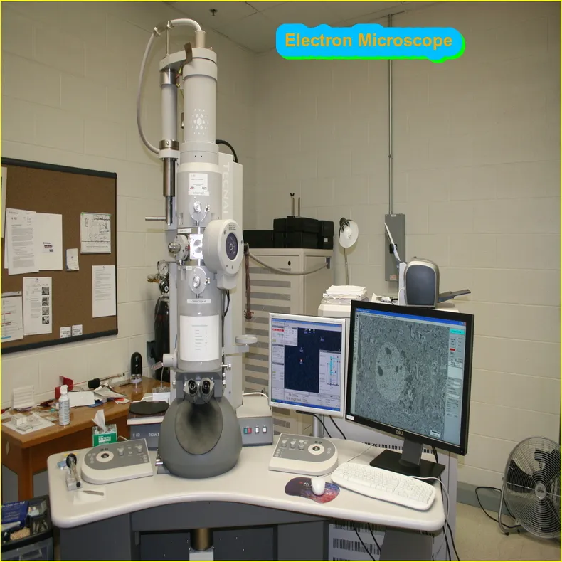
An electrons microscope is a type of microscope that uses a beam of electrons to create an image of the specimen. The first electrons microscope was developed in Germany in 1931 by Ernst Ruska and Max Knoll.
There are two main types of electrons microscopes: transmission electrons microscopes (TEMs) and scanning electron microscopes (SEMs). TEMs use a beam of electrons that passes through a thin specimen to create an image. SEMs use a beam of electrons that scans the surface of the specimen to create an image.
Both TEMs and SEMs have high resolutions, which means they can produce images with great detail. TEMs have a resolution of 0.2 nm, while SEMs have a resolution of 1 nm. Electrons microscopes are used in many different fields, including medicine, biology, and materials science.
Article About:- Health & fitness
Article About:- Medical Technology
Article About:- IR News
Article About:-Amazon Product Review
What is the Principle of Electron Microscope
The principle of an electron microscope is to use a beam of electrons to magnify objects. The electrons are accelerated by an electric field and then passed through a magnetic field, which deflects them onto a screen. The objects being magnified are placed in the path of the electron beam.
The advantage of using an electron microscope over a light microscope is that the wavelength of electrons is much shorter than that of visible light, so they can be used to magnify objects at a much higher resolution. Electron microscopes can also be used to image objects that are too small to be seen with a light microscope, such as viruses and bacteria.
What is the Types of Electron Micropheres
An electron microscope is a type of microscope that uses a beam of electrons to create an image of an object. There are two main types of electron microscopes: transmission electron microscopes (TEMs) and scanning electron microscopes (SEMs).
TEMs work by passing a beam of electrons through a thin specimen, which then produces a corresponding image on a fluorescent screen or photographic film. This type of microscope is typically used for studying the internal structure of materials such as metals, crystals, and biological cells.
SEMs work by bombarding the surface of a specimen with electrons and then observing the resulting interactions between the electrons and the atoms in the specimen. This type of microscope is typically used for studying the external features of objects such as insects, rocks, and plant leaves.
What is the Uses of Electron Microscope
An electron microscope is a powerful tool that allow scientists to see small things at high resolutions. There are two main types of electron microscopes, transmission electron microscopes (TEM) and scanning electron microscopes (SEM). Both types of microscopes use electrons instead of light to create an image.
The major difference between a TEM and SEM is the way in which they create the image. In a TEM, a beam of electrons is passed through a very thin specimen, which then transmits the image onto a detector. In a SEM, the electrons bounce off the surface of the specimen and are detected by an electron detector.
Both transmissions and scanning electrons microscopes can be used to look at a variety of different specimens, including cells, viruses, and minerals. TEMs are typically used to examine very small objects, such as atoms or proteins, while SEMs are better suited for looking at larger objects, such as tissue samples.
While both types of electrons microscopes have their own uses, they also have some limitations. One major limitation is that both TEMs and SEMs require special sample preparation in order for the specimen to be viewed properly. Additionally, because these instruments use electrons instead of light, they can only be used in vacuum chambers.
Scanning electron microscope
A scanning electron microscope (SEM) is a type of electron microscope that produces images of a sample by scanning the surface with a focused beam of electrons. The electrons interact with the atoms in the sample, producing signals that contain information about the surface topography and composition of the sample.
SEMs are capable of achieving high resolutions, due to their use of electrons instead of light waves. They also have a large depth of field, which allows them to image large samples in three dimensions.
SEMs are used in a variety of fields, including materials science, nanotechnology, semiconductor manufacturing, and biology.
Application of electron microscope
An electrons microscope is a type of microscope that uses a beam of electrons to create an image of the specimen. The first electrons microscope was built in 1931 by German physicist Ernst Ruska and his team.
Today, electron microscope are used in a variety of fields, including medicine, materials science, and nanotechnology. They can be used to examine objects at the atomic level and have resolutions that are orders of magnitude higher than those of light microscopes.
There are two main types of electrons microscopes: transmission electron microscopes (TEMs) and scanning electrons microscopes (SEMs). TEMs use a beam of electrons that passes through a thin specimen to create an image. SEMs bounced focused beams of electrons off the surface of the specimen to create an image.
Both types of microscopes have their own advantages and disadvantages. TEMs can provide higher resolutions but are more difficult to use. SEMs are easier to use but have lower resolutions.
Electrons microscopes can be used for a variety of applications, such as studying the structure of cells, examining minerals and metals, and looking at the surfaces of materials.


