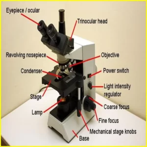
What are the Compound Microscope Parts
Have you ever wanted to take a closer look at the world around you? A compound microscope is a powerful tool that enables us to do just that. In this article, we’ll break down the different parts of a compound microscope and explore their different functions. We’ll also discuss how these parts work together to magnify smaller samples to study in more detail.
What is a compound microscope?
A compound microscope is a type of optical microscope that uses two or more lenses to magnify an object. The first lens, the objective lens, is positioned near the specimen and magnifies it. The second lens, the ocular lens, is located near the eye and magnifies the image further.
Compound microscopes or compound microscope parts are generally more powerful than simple microscopes, as they can achieve greater magnification. However, they are also more complex and sensitive, so more care is required when handling them.
Article About:- Health & fitness
Article About:- Medical Technology
Article About:- IR News
Article About:-Amazon Product Review

Compound microscopes parts and functions
The compound microscope is one of the most important tools in scientific research. It is used to magnify small objects so that they can be studied in detail. A compound microscope consists of several parts, each with a specific function.
The body of the microscope is the largest part and houses all the other parts. The base is attached to the body and provides support for the microscope. The arm extends from the base and supports the objective lenses. The stage is located in the middle of the body and holds the specimen. The stage may also have a movable plate that allows fine adjustment of the position of the specimen. The condenser lens is located below the stage and helps to focus the light onto the sample. The eyepiece lens is located above the stage and magnifies the image of the specimen.
The compound microscope is a powerful tool that allows scientists to study small objects in great detail. By understanding the function of each part, scientists can use the instrument to its full potential.
The microscope and its parts
A compound microscope is a type of optical microscope that uses two or more lenses to magnify an object. The first lens, the objective lens, is placed close to the object being viewed. A second lens, the eyepiece, is placed near the observer’s eye.
The objective lens is usually composed of several separate lenses that work together to magnify the image. The eyepiece also contains multiple lenses, but its main purpose is to magnify the image formed by the objective lens.
A compound microscope parts has three main parts: the body, the stage, and the light source. The body holds all of the lenses in place and provides a way to focus on an object. The stage is where the object being viewed is placed. The light source illuminates the object so that it can be viewed through a microscope.
Compound microscopes can be used for a variety of different purposes, including studying cells, viewing bacteria, and plant life. They are an essential tool for many scientists and researchers.
The light source in a compound microscope
The light source in a compound microscope is usually a halogen lamp, which provides good intensity and color. Light is focused onto the specimen by the condenser lens, and then passes through the objective lens before reaching the eyepiece. To obtain the best possible image, it is important to adjust the light source so that it is perpendicular to the optical axis of the microscope.
How the light travels through the microscope
Light is essential for using a compound microscope. compound microscope parts The vital light source is placed under the stage, and the light travels through the condenser lens before passing through the sample on the stage. The light then passes through the objective lens before reaching the ocular lens (eyepiece).
Most microscopes have an adjustable diaphragm that controls the amount of light passing through the condenser. By adjusting the diaphragm, you can optimize the amount of light that passes through your sample, which will improve image quality.
It is also important to note that different purposes require different amounts of light. For example, a high powered objective will require more light than a low powered objective. If you are having trouble seeing your specimen clearly, try adjusting the diaphragm or changing to a higher-power objective.
The lens in a compound microscope parts
The lens is the most important part of the compound microscope parts. It is responsible for enlarging the sample and making it visible to the user. The compound microscope parts consist of two lenses, the objective lens and the eye lens. The objective lens is positioned near the specimen and is used to project an image of the specimen onto the ocular lens. The ocular lens is located on the eyepiece of the microscope and magnifies the image formed by the objective lens. The two lenses work together to provide high magnification of the specimens. Compound Microscope Parts.


