What is a Slit Lamp ?
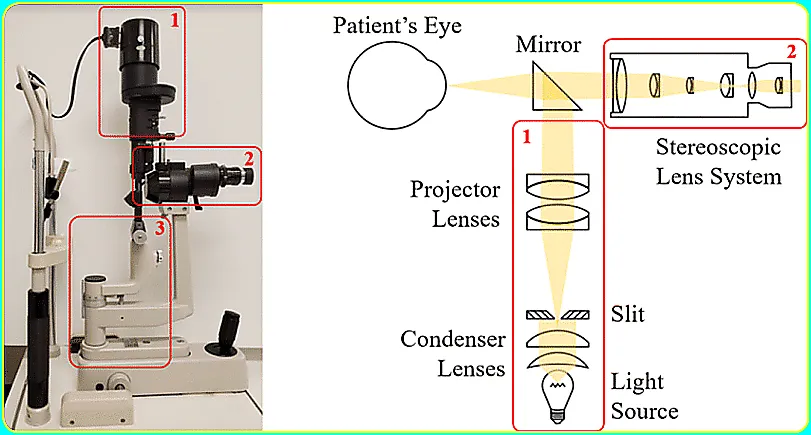
A slit lamp is a microscope in which the eye is examined. Bright light is used during the examination. This helps your ophthalmologist take a closer look at the front of the eye and the various structures inside the eye. It is an important tool in determining the health of your eyes and diagnosing eye disease.
What is a slit lamp test?
Slit lamp examination is a very important standard diagnostic procedure, also known as biomicroscopy. A slit lamp combines a microscope with very bright light allowing the doctor to look into the eye easily.
The slit lamp exam is usually part of a comprehensive eye exam. The person is seated on a chair with his chin and forehead resting on the support, facing the slit lamp.
The ophthalmologist can use this instrument to inspect the eye in detail as well as determine if there are any abnormalities in the eye.
When you go to your doctor, they don’t just check to see if you can read the third line on the eye chart clearly. They are also making sure that your eyes are healthy or not.

Article About:- Health & fitness
Article About:- Medical Technology
Article About:- Soprts
To perform this type of test, many doctors use a device called a “slit lamp.” This is a special microscope and light that lets your doctor see both the inside and outside of your eye in 3-D. They can use it with an ophthalmoscope to see the back of your eye.
Eye diseases are difficult to diagnose during a simple physical exam. A doctor who is an eye doctor who specializes in treating eye problems, called an ophthalmologist, is better able to examine and diagnose these conditions because the equipment they have is specific to the eye. When you have an eye exam, you are more likely to have a slit lamp exam.
Slit lamp examination
What happens during a slit lamp exam?
You do not need to prepare slit lamp in advance for the exam.
When you sit in the exam chair, the doctor will place a device in front of you on which your chin and forehead rest. It helps to keep your head steady for the exam. Your eye doctor may put a few drops in your eye to make any abnormalities on the surface of your cornea more visible. The drops contain a yellow pigment called fluorescein, which washes away your tears. Additional drops may also be put in your eyes so that your pupils dilate, or become larger, to make it easier to see.
Ophthalmologists use a low-powered microscope with a slit lamp, which is a high-intensity light. Slit lamps have different filters to get different views of the eyes so that your eyes can see up close. Some doctors’ offices may have equipment that captures digital images to track changes in the eye over time.
1.) Skin around the eye:- Your doctor may examine the area for skin diseases and abrasions
2.) Your eyelashes and eyelashes:- Styes or oil gland infections, folliculitis or hair follicle infections, and tumors are some of the conditions that your doctor sees.
3.) Eye surface:- This test includes the tissue under your eyelids and above the whites of your eyes. These areas may be inflamed or infected. It can be caused by sexually transmitted diseases, allergies or viruses.
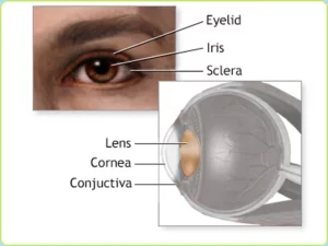
4.) Sclera:- This is the protective outer layer of the eyeball. Next to the sclera is the episclera, which helps to keep it healthy. Allergies, autoimmune disorders, and diseases related to gout or rheumatism can occur in these areas.
5.) Cornea:- This is the lining of the eye which helps in focusing your vision. A slit-lamp exam may show that your cornea is not as clear as it used to be.
6.) The iris:- This is the colored disc, which surrounds the pupil and keeps changing to let more or less light into your eye. It can be affected by a variety of diseases and conditions, including freckles or melanoma of the iris.
7.) Lens:- Cataract is diagnosed by examining this part of the eye. It is located behind the pupil.
What is the process of slit lamp
After taking an initial look at the eyes, the ophthalmologist may apply a special dye called fluorescein to make the examination easier. They administer it as an eye drop or on a small, thin paper strip that touches the white of the eye.
The ophthalmologist will then administer a series of eye drops that dilate the pupils. This dilation makes it easier for the doctor to see other structures in the eye. It takes about 20 minutes for the drops to work.
Once the person has dilated pupils, the doctor will repeat the eye exam. This time they keep a special lens near the eye.
This procedure does not hurt, although there may be some brief prickling during application of the eye drops.
When dilated pupils become too large, which can make the eyes sensitive to light. This can make driving or spending time outdoors uncomfortable. However, the eye drops should be discontinued within a few hours, and wearing sunglasses during this period helps.
What is the working principle of slit lamp
The slit lamp is able to illuminate the tissue of the eye in several different ways, depending on the clinical situation, all of which are useful. The beginner ophthalmologist has to make quick efforts to master all of these illumination techniques, in order to take full advantage of the slit lamp. The slit lamp offers six main illumination options, each of which has its own
Special properties and special uses:

1.) Diffuse Light
2.) Direct Focal Illumination
3.) Specular Reflection
4.) Transillumination, or Retroillumination
5.) Indirect lateral illumination
6.) Sclerotic Scatter
In addition, vibrating the slit beam allows the examiner to observe properties of some ocular tissues that cannot be observed with steady light alone.
What does the slit lamp exam help in the diagnosis?
A slit lamp exam helps diagnose the following conditions:
1.) Macular degeneration, a chronic condition that affects the part of the eye that is responsible for central vision.
2.) A detached retina, a condition when the retina, which is an important layer of tissue at the back of the eye, can detach from its base.
3.) Cataract, a clouding of the lens that negatively affects the ability to see images clearly.
4.) Injury to the cornea, an injury to one of the tissues covering the surface of the eye.
5.) Blockages of retinal vessels, blockages in the blood vessels of the eye that cause sudden or gradual loss of vision.
What to expect after the exam
There are no significant side effects of this test. Your eyes become sensitive to light after a while, especially if your pupils are dilated. If you start to feel nauseous or have eye pain, go back to your doctor’s office as soon as possible and explain this and this problem. These could be symptoms of increased fluid pressure in the eye, which can be a medical emergency. While the risk of this may be small, eye drops used to dilate the eye can rarely cause this to happen.
What does abnormal result mean?
If your slit lamp test results are abnormal, a variety of conditions may be present, including:
1.) Having an infection.
2.) Swelling and burning.
3.) Increased pressure in the eye.
4.) Degeneration of the arteries or veins in the eyes.
For example, if macular degeneration is occurring, the doctor may find drusen, which are yellowish deposits that form in the macula at the onset of age-related macular degeneration. If the ophthalmologist suspects a specific cause of vision problems, they may recommend further testing to obtain a more definitive diagnosis.
How to use slit lamp
Ophthalmologist
For comfortable sitting, move your chair to the right height and adjust the table so that you can sit up straight while looking at the oculars.
Never tilt your head, never tilt your head, never use a slit lamp while standing.
Patient
These will set you up for success, even if it requires you to take a 15-20 minute test. It is important that the patient is comfortable. They are more likely to be able to keep their eyes open, look in the directions you want them to see, and remain still.
1.) Clean, thoroughly clean the application tonometer, chinrest, top strap and handle.
2.) Correctly position the patient’s head, chin in the cup, forehead against the top strap. Line up the patient’s eyes with the markings on the side of the patient stabilization frame.
3.) Tell the patient to “Hold these handles as if you were riding a bicycle.” This slit lamp keeps the table super stable and secure when you are applanating or removing stitches, etc.
4.) Correctly adjust the patient’s chair Move the patient’s chair up or down to adjust the patient’s height.
The best way to move your field of view is not to move the joystick or lens freely. Instead, move the joystick very slowly in the same direction on both the slit lamp and the lens simultaneously. This could be a game changer for many of the people we teach.
How to test cell and flare
Use a 16x mag in which the beam of light is angled from the side. Use bright light in a dark room Look for cells above the pupil This is the darkest part to give you the greatest contrast. Use the following sun grading criteria.
Sun Grading Scheme for Anterior Chamber Cells:-
grade cell in the field
0 < 1
0.5+ 1 – 5
1+ 6 – 15
2+ 16 – 25
3+ 26 – 50
4+ 50+
(using 1mm slit beam)
Sun Grading Scheme for Anterior Chamber Flare:
grade description
0 none
1+ unconscious
2+ medium (iris/lens detail clear)
3+ Marked (Iris/Lens Details Blur)
4+ Acute (fibrin/plastic aqueous)

Article About:- Health & fitness
Article About:- Medical Technology
Article About:- Soprts
Rotate the beam of light about 45 degrees from the center line and move the applanator inline until you feel the click
Adjust the prism so that it is also facing the center line.
Flip your lamp to the blue setting and turn up the brightness all the way. This requires increasing both the beam width and the slit lamp power setting.
Now, look in ocular to make sure the prism is horizontal.
Tilt the control stick backwards. This gives you fine control to move forward and gently descend on the cornea. If you don’t, you can move the lamp around when it’s too close to the cornea. Not only will your patient be scared, but they may close their eyes when you get closer.
Then by tilting the control stick backwards, move within 3-5 mm of the cornea.
Push the control stick forward, using its fine control to move forward and gently land on the corneal surface.
When you do this successfully, you will see the fault, and you will be able to adjust the dial accordingly.
Advantages of slit lamp
1. Having a large field of view.
2. It is easy to check whether the patient has eye movement or not.
3. Useful in the dark because of its bright light and optical property.
4. Through this, abnormalities are diagnosed.
5. Can be used interactively.
Disadvantages of slit lamp
1. Impossible With Very small Pupils
2. More uncomfortable for the patient
3. Low magnification, hence a smaller detail
4. Difficult to learn
How much do slit lamps cost
It usually costs from $450 to $1000. But its price can also change according to the city and region.
Slit lamp biomicroscopy
Due to the small size of the eye segment in this, we need higher magnification and clarity to make finer details. This can be achieved by separating 2 convex lenses at the observer’s end at its focal distance and, for further magnification of the image, a concave-convex combination.
The binocular arrangement of the lens needs to focus near, so a plus power objective lens is fixed. The eyepiece lens inverts the image by default so a Poro-Abe prism is placed in front of it for erect image visualization.
FAQ
How to use a slit-lamp biomicroscopy

Slit-lamp biomicroscopy is a technique used by healthcare professionals, particularly ophthalmologists and optometrists, to examine the anterior and posterior segments of the eye. It provides a highly magnified and detailed view of various eye structures, allowing for the detection and evaluation of eye conditions and diseases.
Preparation.
Adjusting the microscope.
Using the slit beam.
Using the biomicroscope.
What is a slit lamp adapter ophthalmology

In ophthalmology, a slit lamp adapter refers to an accessory or attachment that can be added to a slit lamp biomicroscope. It is designed to enhance the capabilities of the slit lamp by allowing additional diagnostic procedures or specialized examinations to be performed.
How to assemble goldmann tonometer to slit lamp

Assembling a Goldmann tonometer to a slit lamp involves attaching the tonometer head to the microscope of the slit lamp. Here’s a step-by-step guide on how to do it:
Gather the necessary equipment.
Prepare the tonometer prism
Locate the tonometer mounting bracket.
Insert the tonometer head.
Secure the tonometer head.
Attach the tonometer prism.
Align the tonometer prism.
Calibrate the tonometer.
What does the slit lamp test measure

The slit lamp test, also known as biomicroscopy, is a versatile examination technique used in ophthalmology to assess various structures of the eye in detail. It allows healthcare professionals to visualize and evaluate the anterior and posterior segments of the eye.
Anterior Segment Assessment.s.
Anterior Chamber.
Anterior Chamber Angle.
Posterior Segment Assessment.
Tear Film Evaluation.
What is slit-lamp ophthalmoscopy

Slit-lamp ophthalmoscopy, also known as direct ophthalmoscopy or biomicroscopic ophthalmoscopy, is a technique used to examine the posterior segment of the eye, specifically the retina, optic nerve, and blood vessels. It involves using a slit lamp biomicroscope along with specialized lenses or attachments to provide a magnified view of the interior structures of the eye.
Preparation.
Set the appropriate magnification and illumination.
Patient positioning.
Performing slit-lamp ophthalmoscopy.


