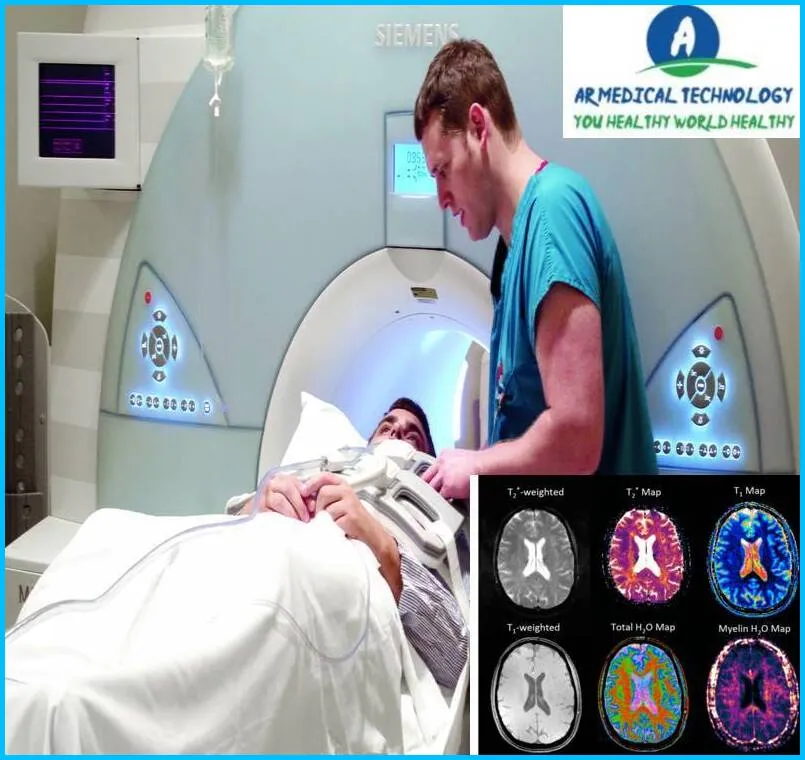
Normal Brain MRI with Contrast, Normal Brain MRI with Contrast
Normal Brain MRI with Contrast
Normal Brain MRI with Contrast: What is a brain MRI with contrast exactly? Certain brain MRI studies require an injection of a contrast material. The rare earth element gadolinium is commonly used as the contrast agent. This substance alters the magnetic characteristics of nearby water molecules when it is present in your body, which enhances the quality of the image.
Will you be undergoing a regular contrast-enhanced brain MRI? If so, you’ve arrived to the right place! Strong magnets and radio waves are used in magnetic resonance imaging (MRI), a non-invasive diagnostic technique that produces detailed images of your brain. Finding any anomalies or damage inside the brain might be facilitated by a standard brain MRI scan.
When paired with contrast dye, it produces significantly more detailed pictures and aids in the identification of medical issues that may go undetected on standard scans. In this blog article, we’ll go over what a normal brain MRI scan with contrast entails and why it’s such an important diagnostic tool for many medical professionals. So let’s get this party started.
Normal MRI Brain Scan Vs Abnormal
Different disorders or anomalies in the structure and function of the brain can be revealed by an MRI (Magnetic Resonance Imaging) scan, which can also be performed abnormally. An outline of what may be seen in each scenario is provided below:
Normal MRI Brain Scan:
- Clear Structures:
- A normal MRI brain scan will display clear and well-defined structures of the brain, including the cerebral cortex, white matter, and various subcortical structures.
- Symmetry:
- Both hemispheres of the brain should appear symmetrical in shape and size. Any significant asymmetry may warrant further investigation.
- Ventricles:
- The ventricular system, which includes the lateral ventricles, third ventricle, and fourth ventricle, should appear normal in size and shape.
- Absence of Lesions:
- There should be an absence of abnormal growths, tumors, cysts, or lesions in different regions of the brain.
- Blood Vessels:
- Blood vessels, including arteries and veins, should be clearly visible and exhibit normal flow patterns.
- No Signs of Bleeding:
- There should be no evidence of bleeding or hemorrhage within the brain tissue.
Abnormal MRI Brain Scan:
- Tumors or Lesions:
- Abnormal brain scans may reveal the presence of tumors, cysts, or other growths that can disrupt the normal brain structure.
- Brain Atrophy:
- Atrophy or shrinkage of certain brain regions may be indicative of neurodegenerative conditions such as Alzheimer’s disease.
- White Matter Changes:
- Changes in the appearance of white matter can indicate conditions such as multiple sclerosis or other demyelinating disorders.
- Stroke or Ischemia:
- Evidence of stroke, ischemia, or infarction may be visible as areas of abnormal signal intensity or tissue damage.
- Vascular Abnormalities:
- Abnormalities in blood vessels, such as aneurysms or arteriovenous malformations, may be detected.
- Inflammation or Infection:
- Conditions involving inflammation or infection, such as encephalitis or meningitis, may be evident in abnormal MRI findings.
- Hydrocephalus:
- Enlargement of the ventricles may indicate conditions like hydrocephalus, where there is an abnormal accumulation of cerebrospinal fluid.
- Trauma:
- Evidence of trauma, such as contusions or bleeding, may be visible in cases of head injury.
A normal MRI brain scan should reveal no abnormalities or damage. However, if there is an issue with the brain, it may appear as a mass, tumor, cyst, or other abnormality on the scan. An MRI may also be performed in some circumstances to search for signs of a stroke or other disorders.
Article About:- Health & fitness
Article About:- Medical Technology
Article About:- Sports

Normal Brain MRI side view
A normal brain MRI side view should show the cerebrum, cerebellum, and brainstem. The cerebrum is the large, wrinkly part of the brain that controls voluntary movement, speech, thought, and emotion. The cerebellum is the small, smooth part of the brain that helps control balance and coordination. The brainstem is the narrow stalk of tissue that connects the cerebrum to the spinal cord.
Normal brain MRI with labels
A normal brain MRI with labels is a great way to look at different parts of the brain and how they work together. The labels can help you understand what you’re looking at and how the different parts of the brain interact.
Normal Brain MRI Report Sample
Brain MRI with contrast is a test that uses magnetic resonance imaging (MRI) to produce detailed images of the brain. The test is also called MRI of the brain with contrast, cerebral MRI with contrast, or brain scan with contrast.
The MRI machine creates a strong magnetic field around the patient’s head. The magnetic field aligns the hydrogen atoms in the body. Radio waves are then sent through the body to jolt the hydrogen atoms out of alignment. As the atoms return to their original positions, they emit signals that are picked up by the receiver in the MRI machine.
The computer processes these signals and produces a picture on the monitor. The images show structures in the brain, including neurons (nerve cells), blood vessels, and other tissues. Images can be viewed from different angles and can be magnified.
Contrast agent is injected into a vein before the MRI procedure begins. The contrast agent contains gadolinium, which helps to improve the quality of the images by making certain structures more visible.
How to Read MRI Brain Stroke
If you or a loved one have recently had a brain stroke, you may be wondering how to read an MRI brain scan. Here, we’ll provide a brief overview of what you can expect to see on an MRI brain scan of a stroke victim.
First, it’s important to note that there are two types of stroke: hemorrhagic and ischemic. Ischemic stroke occurs when blood flow to the brain is blocked, while hemorrhagic stroke occurs when a blood vessel in the brain bursts. Each type of stroke will show up differently on an MRI brain scan.

Ischemic strokes usually show up on the scan as a dark area, known as an infarct. This is because the lack of blood flow causes cell death in that area of the brain. On the other hand, a hemorrhagic stroke will appear as a bright spot on the scan due to the presence of blood in the brain tissue.
Depending on the location and severity of the stroke, other changes may be seen on an MRI brain scan. For example, if a stroke has caused damage to the white matter of the brain, this will show up as areas of high signal intensity (called leukoaraiosis). Swelling or inflammation in the brain can also be seen as increased signal intensity in an MRI scan.
In some cases, it can be difficult to determine whether a person has had a stroke based on an MRI brain scan alone.
How to Read MRI Brain Tumor
A specific expertise of neuroimaging is necessary in order to interpret an MRI (Magnetic Resonance Imaging) scan for brain malignancies. Here is a broad advice on how to interpret an MRI brain scan for potential malignancies, albeit a precise diagnosis can only be made by a competent healthcare practitioner, such as a radiologist or neurologist:
Reviewing the Images:
- Examine the series of images obtained from different angles and sections of the brain. MRI scans typically consist of various sequences like T1-weighted, T2-weighted, and contrast-enhanced images.
2. Identifying Normal Structures:
- Familiarize yourself with normal brain anatomy. Understand the appearance of structures such as the cerebral cortex, white matter, gray matter, ventricles, and major blood vessels.
3. Looking for Abnormalities:
- Pay attention to any abnormal growths, masses, or lesions within the brain tissue. Tumors can vary in size, location, and characteristics on MRI.
4. Characteristics of Tumors:
- Tumors on MRI scans may appear as areas of abnormal signal intensity compared to the surrounding healthy tissue.
- Features such as shape, borders, and enhancement patterns (with contrast) can provide clues about the nature of the tumor.
5. Differentiating Types of Tumors:
- Gliomas (brain tumors arising from glial cells) may show variable signal intensities on both T1 and T2-weighted images.
- Metastatic tumors (those spreading from other parts of the body) may have a characteristic appearance, often with ring-enhancement patterns.
6. Assessing Surrounding Structures:
- Evaluate how the tumor interacts with adjacent structures. Tumors may compress nearby tissues, cause edema (swelling), or lead to displacement of surrounding structures.
7. Contrast Enhancement:
- Contrast agents, such as gadolinium, are often used to enhance the visibility of tumors. Enhanced areas on MRI may indicate increased blood flow or breakdown of the blood-brain barrier, which is common in tumors.
8. Necrosis and Cystic Components:
- Look for regions of necrosis (dead tissue) within the tumor, which may appear as dark areas on imaging. Cystic components or fluid-filled spaces may also be present in certain tumors.
9. Size and Growth:
- Assess the size and location of the tumor. Changes in size or growth patterns over time may be important for diagnosis and treatment planning.
10. Consultation with Specialists:
- It’s crucial to involve specialists, such as neurosurgeons or oncologists, in the interpretation process. A multidisciplinary approach is often necessary for accurate diagnosis and treatment planning.
MRI brain tumors can be very difficult to read, but there are some things you can do to make it easier. First, you need to understand what an MRI brain tumor is. MRI brain tumor is a mass or lesion that is visible on an MRI scan of the brain. It is important to note that not all tumors are cancerous, and many benign tumors do not cause any symptoms. However, if you have a brain tumour, it is important to see a doctor so they can determine if it is cancer.
There are two types of contrast used in MRI: T1 and T2. T1 contrast helps to show areas of high cell density, while T2 contrast helps to show areas of low cell density. When looking at an MRI scan of the brain, you will want to look for any areas of abnormality. These areas may be darker or lighter than the surrounding tissue, and they may differ in size from the rest of the brain. If you notice any areas that look abnormal, you should speak to your doctor so they can investigate the issue further.

Frequently Asked Questions
What is a brain MRI with contrast looking for?

Brain. If you’ve been in a car accident and suffered a brain injury, an MRI with contrast will reveal your injury in better detail than an MRI without contrast. It can also detect brain cancers and aid in the diagnosis of multiple sclerosis, stroke, dementia, and a brain infection.
What is the difference between normal and contrast MRI brain?

A contrast agent is used in contrast MRIs, whereas non-contrast MRIs do not. As a result, additional follow-up procedures to clarify anomalies may be required. Non-contrast MRIs are more difficult to interpret than contrast MRIs.
Do you really need contrast for brain MRI?

Contrast dye is not required for the majority of MRI services. If a non-contrast MRI does not generate a clear enough image, an extra contrast MRI may be needed. Because air and hard bone do not emit an MRI signal, they look black or very dark on the image.
What abnormalities can be seen on a brain MRI?

Abnormal outcomes might be caused by: abnormal blood arteries in the brain (head arteriovenous malformations)
Acoustic neuroma is a tumor of the nerve that links the ear to the brain.
There is bleeding in the brain.
Infection of the brain.
Brain tissue enlargement.
Tumors of the brain.
An injury to the brain causes brain damage.
Can MRI with contrast detect brain tumor?

Gliomas are among the most frequent types of brain tumors. Gliomas are often evaluated with contrast enhanced MRI, which has a high sensitivity, moderate specificity, and good diagnostic accuracy in detecting intra-axial gliomas.
Why would a neurologist order a brain MRI with contrast?

Contrast is frequently used to improve the visibility of blood arteries and tumors, as well as to identify stroke, infection, multiple sclerosis, and dementia. If contrast is requested for your MRI, the technician will inject it into a vein in your hand or arm prior to taking pictures.


