Multiple Sclerosis Normal Vs MS Brain MRI Images
Are you curious about the difference between a normal brain MRI and one from multiple sclerosis (MS)? look no further! In this blog post, we’ll explore the striking contrasts in imaging that accompany MS. By examining these images side by side, you will gain a deeper understanding of what is happening inside the brains of people who suffer from the condition. Let’s dive into the details and see how technology is helping us better understand MS.
Normal vs MS brain MRI images
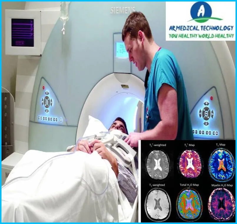
When it comes to brain MRI images, there are some important differences between a normal brain and a brain affected by multiple sclerosis (MS). First, let’s take a look at a typical brain MRI. The image will show different parts of the brain, including:
-Cerebral cortex
-Subcortical areas
-Basal ganglia
-Thalamus
-Cerebellum
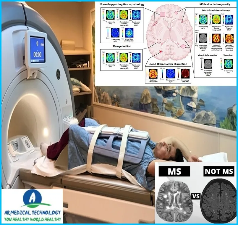
Now, let’s compare that to an MRI of a brain affected by MS. There are a few key differences:
- There may be lesions or plaques in the white matter of the brain.
- The lesions may be in different stages of development, from early to advanced.
- The size and number of lesions can vary from person to person.
So, what do these differences mean? Essentially, MS affects the white matter of the brain, which is responsible for communication between different areas of the brain. The lesions cause disruptions in this communication, leading to the symptoms of MS.
Article About:- Health & fitness
Article About:- Medical Technology
Article About:- IR News
Article About:- Sports

Multiple sclerosis MRI vs normal
Multiple sclerosis (MS) is a progressive neurological disorder that attacks the central nervous system (CNS), which includes the brain, spinal cord and optic nerves. The damage caused by MS can lead to a variety of symptoms, including problems with vision, movement, sensation, and balance.
MRI is the best tool for diagnosing MS because it can show lesions in the CNS that are characteristic of the disease. Normal brain MRI images do not show these lesions.
Comparing an MRI of someone with MS to a normal brain MRI can help doctors confirm a diagnosis of MS and rule out other possible conditions. It can also help them determine the severity of the disease and track its progress over time.
Does early MS show up on MRI
Multiple sclerosis (MS) is a chronic autoimmune disease that attacks the central nervous system (CNS), which includes the brain, spinal cord, and optic nerves. Early MS symptoms may include fatigue, bowel and bladder problems, numbness or tingling in the limbs, vision problems, and weakness. Many of these symptoms can also be caused by other conditions, so it can be difficult to diagnose MS initially.
MRI is one tool that can be used to help diagnose MS. An MRI of the brain can show the lesions or areas of damage that are characteristic of MS. However, not everyone with MS will have detectable lesions on MRI, especially in the early stages of the disease. In addition, some people with other neurological conditions may also have brain lesions that may look similar to those seen in MS on MRI. Therefore, MRI alone is not sufficient to diagnose MS. Other factors, such as a person’s symptoms and medical history, must also be considered when making a diagnosis.
What do, MS Lesions Look Like on MRI
There are several ways that lesions, or damage, from MS can show up on an MRI. The most common type of lesion is called a T2 hyperintense lesion. These lesions appear as bright spots on an MRI and are thought to be caused by inflammation and/or damage to the myelin. Other types of lesions include T1 hyperintense lesions (which appear as dark spots on MRI), FLAIR hyperintense lesions, and gadolinium-enhancing lesions. The lesions may vary in size and number, and may be located in different parts of the brain and/or spinal cord.
MS brain MRI Without Contrast
Normal brain MRI vs MS brain MRI (without contrast)
A typical brain MRI shows the structures of the brain and spinal cord in great detail. This can help doctors diagnose problems such as stroke, tumors, aneurysms, and other conditions.
MS is a disease that attacks the central nervous system, which includes the brain and spinal cord. An MRI of the brain can show the lesions, or areas of damage, that are characteristic of MS. These lesions may be small and scattered or large and clustered together.
MS is a diagnosis made based on clinical symptoms and findings from an MRI. In some cases, a contrast agent may be used during an MRI to help highlight abnormalities. However, an MRI of the brain can often be diagnostic even without contrast.


