What is the Molar Pregnancy Ultrasound
Molar Pregnancy Ultrasound is a condition that can occur when a fertilized egg doesn’t develop properly. Instead of developing into a baby, the fertilized egg forms an abnormal mass of tissue. This mass of tissue is called a molar pregnancy. Molar pregnancy can cause serious complications, so it’s important to be aware of the symptoms and seek medical treatment if you suspect you have a Molar Pregnancy Ultrasound.
Molar Pregnancy Ultrasound
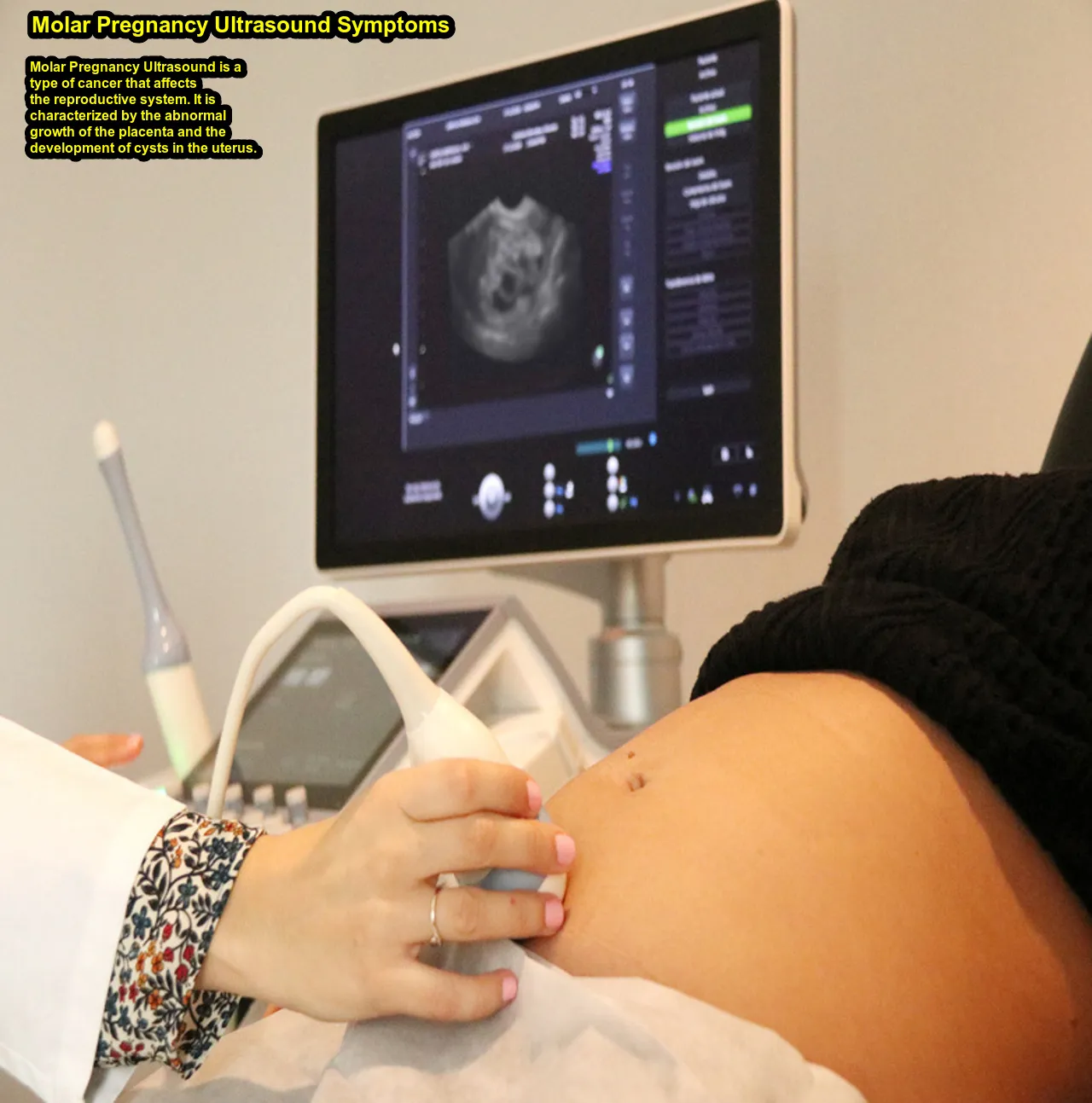
Molar Pregnancy Ultrasound is a type of cancer that affects the reproductive system. It is characterized by the abnormal growth of the placenta and the development of cysts in the uterus. Molar pregnancy can be diagnosed through ultrasound. The most common symptom of molar pregnancy is vaginal bleeding. Other symptoms include abdominal pain, nausea, and vomiting. Molar Pregnancy Ultrasound can be treated with surgery or chemotherapy.
A molar pregnancy is a rare complication of pregnancy in which an abnormal mass of tissue develops in the uterus. This mass, known as a mole or gestational trophoblastic disease (GTD), forms instead of a normal pregnancy. A molar pregnancy can be partial or complete, and it often requires medical intervention.
Role of Ultrasound in Detecting Molar Pregnancy:
- Early Detection: Ultrasound is a key tool in early pregnancy monitoring. In the case of a molar pregnancy, an ultrasound can help detect abnormalities in the development of the gestational sac and the absence of a viable fetus.
- Abnormal Appearance: A molar pregnancy may appear as a grape-like cluster of cystic structures within the uterus. The ultrasound can reveal the characteristic vesicles or clusters associated with molar pregnancies.
- Size of the Uterus: Molar pregnancies can cause the uterus to be larger than expected for the gestational age. An ultrasound helps assess the size of the uterus and the abnormal growth pattern.
- Hydatidiform Mole Identification: There are two main types of molar pregnancies: complete and partial. Ultrasound can help differentiate between these types based on the appearance of the tissue and the presence or absence of a fetal component.
- Follow-up Monitoring: After the evacuation of a molar pregnancy, ultrasounds are often used for follow-up monitoring to ensure that no residual tissue remains in the uterus. This is important to prevent complications such as persistent gestational trophoblastic disease.
Importance of Diagnosis Molar Pregnancy Ultrasound:
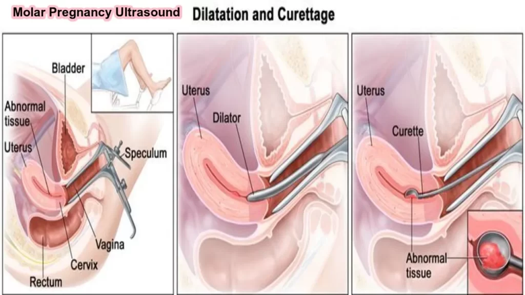
- Treatment Planning: A molar pregnancy requires prompt medical attention. Diagnosis through ultrasound helps healthcare providers plan appropriate treatment, which often involves a procedure called dilation and curettage (D&C) to remove the abnormal tissue.
- Prevention of Complications: Early detection and management of molar pregnancies are crucial to prevent potential complications, such as persistent trophoblastic disease or the development of choriocarcinoma (a type of cancer).
- Emotional Support: The diagnosis of a molar pregnancy can be emotionally challenging for the parents. Ultrasound findings, along with counseling from healthcare providers, can provide information and support during this difficult time.
Molar Pregnancy Ultrasound can cause serious complications for both the mother and the baby. Molar pregnancy can lead to preterm labor and delivery, low birth weight, and stillbirth. Molar pregnancy can also cause problems with the placenta, such as placenta previa (when the placenta covers the cervix) and placenta accrete (when the placenta grows into or through the uterine wall). Molar pregnancy can also cause bleeding during pregnancy and after delivery. Molar Pregnancy Ultrasound is treated with surgery to remove the abnormal tissue. Surgery is usually successful in treating molar pregnancy. However, molar pregnancy can come back after treatment.
Article About:- Health & fitness
Article About:- Medical Technolo
Article About:- Sports

5 Week Twin Pregnancy Ultrasound
Around week six or seven of a normal pregnancy, you would expect to see a yolk sac and fetal pole during an ultrasound scan. But with a molar pregnancy, the ultrasound picture looks very different. Instead of a fetus, you will see what looks like a large, dark mass in the uterus. This mass is made up of abnormal tissue that grows in place of a baby. Molar pregnancies are also known as gestational trophoblastic disease (GTD).
While molar pregnancies are rare, they’re not unheard of. In fact, it’s estimated that 1 in every 1,200 to 1,500 pregnancies is a molar pregnancy. And women who have had one molar pregnancy have about a 1 in 10 chance of having another one. There are two types of molar pregnancies: complete and partial. A complete molar pregnancy is when all of the placental tissue is abnormal. With a partial molar pregnancy, there’s both abnormal and normal tissue present.
Abnormal tissue growth can cause the uterine walls to thin and rupture, which can be life-threatening for both mother and baby. For this reason, it’s important to get regular ultrasounds during a molar pregnancy to check on the status of the abnormal tissue growth and make sure that the uterine walls are still intact. Molar Pregnancy Ultrasound, Molar Pregnancy Ultrasound.
9 week ectopic pregnancy ultrasound
If you are having a Molar Pregnancy Ultrasound, your ultrasound will likely show an empty gestational sac. The gestational sac is the structure in the uterus that holds the developing baby. If you have a molar pregnancy, it means that there is no baby developing inside of the gestational sac. Instead, you will see abnormal tissue growth inside of the sac. This tissue growth can take on a variety of different appearances, depending on the type of molar pregnancy that you have.
If you have a complete molar pregnancy, the gestational sac will be filled with grape-like clusters of tissue. This tissue is not normal pregnancy tissue. Instead, it is made up of abnormal cells that grow quickly and out of control.
If you have a partial molar pregnancy, the gestational sac will contain both normal pregnancy tissue and abnormal tissue. The abnormal tissue will again take on a grape-like cluster appearance. Molar Pregnancy Ultrasound, Molar Pregnancy Ultrasound.

Signs of Down Syndrome During Pregnancy Ultrasound
There are several signs of Down syndrome that can be seen on an ultrasound during pregnancy. The most common is an abnormal shape of the fetus’s head, called a “micro brachycephaly.” This is usually accompanied by an enlarged fluid-filled space in the fetal brain called the ventricles. Other signs include a thickened nuchal fold (the skin at the back of the neck), shortening of the bones in the forearm, and poor development of ossification centers in the bones. These findings are often subtle, and not all fetuses with Down syndrome will have all of them.
What is the Procedure of Molar Pregnancy Ultrasound
The procedure for a molar pregnancy ultrasound is similar to a routine prenatal ultrasound, but with a focus on detecting and assessing the specific characteristics associated with molar pregnancies. Here is an overview of the procedure:
1. Scheduling the Ultrasound:
- The ultrasound may be scheduled if there are signs or symptoms that raise suspicion of a molar pregnancy, such as abnormal bleeding, enlarged uterus, or elevated levels of human chorionic gonadotropin (hCG) in the blood.
2. Preparation:
- Generally, there is minimal preparation for a molar pregnancy ultrasound. The patient may be asked to have a full bladder, as this can improve the visibility of the pelvic structures.
3. Positioning:
- The patient will lie down on an examination table. For a transabdominal ultrasound, a clear gel is applied to the abdominal area. For a transvaginal ultrasound, a specialized transducer is covered with a sterile sheath and inserted into the vagina.
4. Transducer Movement:
- The ultrasound technologist, also known as a sonographer, will move the transducer over the abdominal or pelvic area to obtain images of the uterus.
5. Image Acquisition:
- The ultrasound machine emits high-frequency sound waves that bounce off structures in the pelvic region. The returning echoes are then converted into real-time images on a monitor.
6. Visualization of Uterine Structures:
- The ultrasound allows visualization of the uterus, gestational sac, and surrounding structures. In the case of a suspected molar pregnancy, the sonographer will look for characteristic features, such as the presence of grape-like clusters or vesicles.
7. Differentiation Between Complete and Partial Mole:
- A molar pregnancy can be classified as complete or partial. Complete moles typically show a characteristic appearance on ultrasound, including a snowstorm or vesicular pattern. Partial moles may have more recognizable fetal structures.
8. Measurement of Uterine Size:
- The size of the uterus is assessed to determine if it is larger than expected for the gestational age. Molar pregnancies often lead to abnormal growth patterns.
9. Doppler Ultrasound (Optional):
- Doppler ultrasound, which assesses blood flow, may be used to evaluate the blood vessels within the uterus. This can provide additional information about the nature of the molar pregnancy.
10. Documentation and Reporting:
- The images obtained during the ultrasound are documented, and a report is generated. The report may include findings related to the suspected molar pregnancy and may guide further medical management.

FAQ
How does a molar pregnancy appear on ultrasound?

During a molar pregnancy, the scan may show a characteristic ‘snowstorm’ appearance. You will also find that there is no foetal tissue or only partial foetal tissue. If your blood tests or ultrasound indicate a molar pregnancy, your midwife will inform you. It can be very upsetting to experience this.
Can you tell a molar pregnancy from ultrasound?

In the case of molar pregnancy, ultrasound is often used to identify a complex vesicular intrauterine mass containing many ‘grape-like’ cysts.
What are the hCG levels for molar pregnancy?

A complete mole may result in an increased serum beta-hCG level, often exceeding 100,000 IU/L in women. In case of a partial mole, the beta-hCG level is usually within the wide range associated with normal pregnancy and the symptoms are usually less pronounced.
When do molar pregnancy symptoms start?

A woman with a molar pregnancy is more likely to pass blood clots or have a brownish vaginal discharge. Some women pass molar tissue in the form of small bunches of grapes. It usually begins between weeks 6 and 12 of pregnancy that women begin bleeding from a molar pregnancy.


