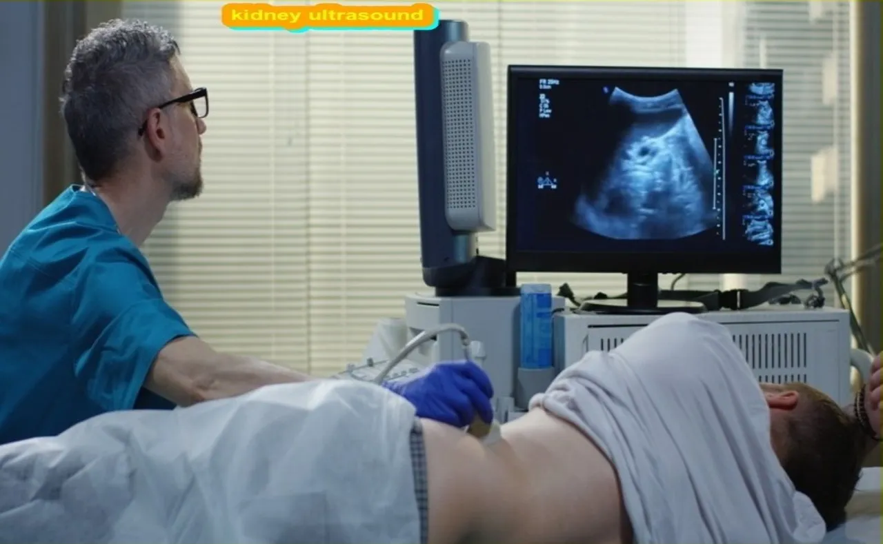
What is the Kidney Ultrasound Procedure
Kidney ultrasound is a non-invasive diagnostic procedure used to assess the health of your kidneys. Ultrasound waves are used to create images of the kidneys, which can be reviewed by the doctor to look for any abnormalities.
This procedure is generally quick and painless, and does not require any special preparation beforehand. However, you may be asked to drink plenty of fluids so that your kidneys are fully hydrated and easier to see on the ultrasound. If you have any pre-existing diseases, please make sure to tell your doctor before the procedure so that they can take proper precautions.
Renal ultrasonography, another name for kidney ultrasound, is a type of medical imaging in which pictures of the kidneys and their surrounding tissues are produced using high-frequency sound waves. This non-invasive imaging method aids medical practitioners in determining the kidneys’ dimensions, form, and state. An outline of the renal ultrasonography process is provided below:
Preparation:
- Generally, there is minimal preparation required for a kidney ultrasound. In most cases, you won’t need to fast or make any special dietary changes.
- It’s advisable to wear comfortable and loose-fitting clothing, as you may be asked to remove certain items for the examination.
2. Patient Positioning:
- You will typically lie on your back on an examination table.
- A clear, water-based gel is applied to the skin over the area where the kidneys are located. This gel helps to transmit the sound waves and improves the quality of the ultrasound images.
3. Ultrasound Transducer:
- The ultrasound technologist, or sonographer, uses a handheld device called a transducer. The transducer emits high-frequency sound waves and captures the echoes as they bounce back from the internal structures.
4. Scanning Procedure:
- The sonographer moves the transducer across the skin surface, directing the sound waves toward the kidneys.
- The sound waves are then converted into images that appear on a monitor in real-time.
- The sonographer may ask you to change positions or take deep breaths to obtain different views and enhance image quality.
5. Evaluation of Kidneys and Surrounding Structures:
- The ultrasound allows visualization of the kidneys, ureters, and bladder.
- The sonographer examines the size, shape, and texture of the kidneys, as well as the presence of any abnormalities such as cysts, stones, or tumors.
6. Completion and Results:
- Once the necessary images have been obtained, the gel is wiped off, and the procedure is complete.
- The results are typically provided to a radiologist, who interprets the images and generates a report for the referring healthcare provider.
Kidney Ultrasound
The kidneys are a pair of organs located in the lower abdominal cavity. They are responsible for filtering waste products from the blood and excreting them in the urine.
Ultrasound is a non-invasive, painless test that does not use ionizing radiation. It is often used to evaluate the size, shape, and location of the kidneys. It can also be used to assess blood flow through the kidneys and to detect abnormalities such as cysts, tumors, or stones.
This entire procedure is usually done in an outpatient setting. You will lie on your back on an exam table with your knees bent. A gel will be applied to your skin to help reduce friction between the transducer and your body. The transducer will then be placed on your abdomen and moved around until it gets a clear view of the kidney.
1. Purpose:
- The primary purpose of a kidney ultrasound is to assess the anatomy and function of the kidneys. It can help identify abnormalities, such as kidney stones, cysts, tumors, or structural issues.
2. Preparation:
- In most cases, little to no preparation is required. However, the healthcare provider may ask you to drink plenty of water before the test to fill the bladder, which helps provide better visualization of certain structures.
3. Patient Positioning:
- You will typically lie on your back on an examination table. The healthcare provider may use pillows or cushions to help position you for optimal imaging.
4. Application of Gel:
- A clear, water-based gel is applied to the skin over the area where the kidneys are located. This gel helps transmit sound waves and improves the quality of the ultrasound images.
5. Ultrasound Transducer:
- The ultrasound technologist, also known as a sonographer, uses a handheld device called a transducer. This device emits high-frequency sound waves into the body and captures the echoes as they bounce back.
6. Image Acquisition:
- The sonographer moves the transducer across the skin surface, directing the sound waves toward the kidneys. The echoes are converted into real-time images on a monitor.
7. Evaluation of Kidneys and Surrounding Structures:
- The sonographer evaluates the size, shape, and texture of the kidneys. The ultrasound can also visualize the ureters, bladder, and surrounding abdominal structures.
8. Completion and Results:
- Once the necessary images have been obtained, the gel is wiped off, and the procedure is complete.
- The ultrasound images are interpreted by a radiologist, who generates a report for the referring healthcare provider.
Diagnostic and surveillance of kidney diseases, such as kidney stones, cysts, infections, and congenital anomalies, are frequently accomplished through the use of renal ultrasound technology. Providing real-time information without using ionizing radiation, it is an invaluable imaging tool. A renal ultrasound may be suggested by your healthcare practitioner as part of the diagnostic procedure if you have symptoms like discomfort or changes in urine or if you have concerns about the health of your kidneys.
Article About:- Health & fitness
Article About:- Medical Technology
Article About:-Sports

Abnormal Kidney Ultrasound
Renal ultrasound is performed to evaluate the size, shape, and location of the kidneys and to assess blood flow through the kidneys. The exam can help detect masses, cysts, stones, or other abnormalities.
A normal kidney ultrasound should show two kidneys that are approximately the same size and shape. The renal arteries and veins should be visible and there should be no obstruction to blood flow. An abnormal kidney ultrasound may show one or more of the following: an enlarged or small kidney, a mass or cyst, a urinary tract obstruction, abnormal blood flow, or calcification.
Normal Kidney Ultrasound
Kidney ultrasound waves are produced by a transducer that is placed on the skin over the area of interest. The waves travel through the body and collide with organs and structures. The echoes are recorded and converted into images that can be viewed on a monitor.
The test is usually performed as an outpatient procedure and does not require any special preparation. You will be asked to drink plenty of fluids before the test to fill your bladder. This helps in improving the quality of the images.
There is no radiation involved in this test and it is considered safe for all patients including pregnant women and children.
Preparation for Kidney Ultrasound
Preparing to have a kidney ultrasound is simple. There is no need for any special preparation to do this. You will be asked to drink plenty of fluids in the days leading up to the test so that your kidneys fill up with fluid. This makes it easier for the ultrasound waves to travel through your body and produce a clear image of your kidneys.
Medullary Sponge Kidney Ultrasound
Medullary sponge kidney (MSK) is a congenital anomaly of the kidney characterized by dilatation and cystic degeneration of the medullary collecting ducts. It is the cause of nephrolithiasis and kidney dysfunction.
Kidneys are vital organs that perform many important functions, including filtering waste products from the blood and excreting them in urine. The kidneys are also involved in regulating blood pressure, electrolyte balance and red blood cell production.
Ultrasound is a painless, non-invasive test that uses sound waves to create images of structures inside the body. Ultrasound can be used to evaluate the size, shape, and location of the kidneys, as well as to detect any abnormalities.
In medullary sponge kidney, the medullary collecting ducts are dilated and cystic. This can lead to nephrolithiasis (kidney stones) and kidney dysfunction.
If you have been diagnosed with medullary sponge kidney or if you have symptoms that may indicate the condition, your doctor may order an ultrasound exam of your kidneys.

Horseshoe Kidney Ultrasound
A horseshoe kidney is a congenital anomaly in which the two kidneys are fused at the lower poles. The shape of the kidney is similar to a horseshoe.
Horseshoe kidney is the most common type of renal fusion anomaly, accounting for approximately 5% of all cases of renal fusion.
Ultrasound is the best method for the initial evaluation of horseshoe kidney. Findings on ultrasound that are suggestive of horseshoe kidney include:
- Ectopic location of the kidneys (i.e., not in the usual anatomical position within the lumbar region)
- Fusion of the lower poles of the kidneys
- Crossing of the renal vessels over the midline (i.e., crossing atypically)
- Duplication of ureters

FAQ
Can an ultrasound detect kidney disease?
Mainly, an ultrasound at the kidneys can examine a patient’s kidney fitness. By using doing so, a health practitioner can determine if there are any present abnormalities in the kidneys. The ultrasound consequences allow them to correctly diagnose kidney ailment—if gift in the body. Moreover, an ultrasound is a non-invasive system.
What are the 3 early warning signs of kidney disease?
Three warning signs and symptoms That you’ll be Experiencing Kidney Failure
Dizziness and Fatigue. One of the first possible signs of weakening kidneys is the revel in of typical weakness in yourself and your basic fitness.
Swelling (Edema)
Adjustments in urination.
Which ultrasound is best for kidney?
A renal (REE-nul) ultrasound makes use of sound waves to make photos of the kidneys, ureters, and bladder. All through the experiment, an ultrasound system sends sound waves into the kidney area and pics are recorded on a laptop. The black-and-white photographs show the inner shape of the kidneys and associated organs.
Can ultrasound detect kidney blockage?
A kidney ultrasound can be used to assess the size, area, and form of the kidneys and associated systems, such as the ureters and bladder. Ultrasound can discover cysts, tumors, abscesses, obstructions, fluid collection, and infection within or around the kidneys.
What creatinine level is Stage 3?
Most fulfilling cutoff values for serum creatinine within the diagnosis of level three CKD in older adults were > or =1.3 mg/dl for guys and > or =1.0 mg/dl for girls, regardless of the presence or absence of high blood pressure, diabetes, or congestive coronary heart failure.


