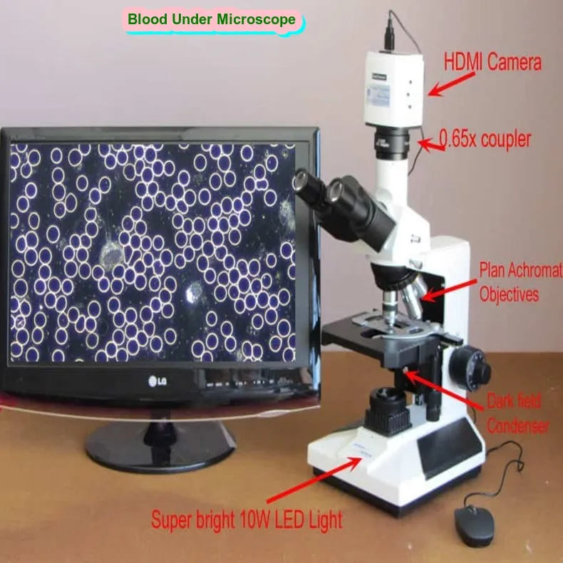
Blood under microscope
Blood Under Microscope What is it
People tend to assume a lot about the world around them, but the reality is frequently different, as demonstrated by the hue of blood in Blood Under the Microscope. In this blog post, we’ll examine and disprove a few “facts” that our culture takes for granted, such as the definition of “red,” the number of blood cells that qualify as anemia, and what blood looks like under a microscope.
Why is blood red?
There are a few explanations for why blood appears red in Blood Under Microscope. Hemoglobin, a substance in red blood cells that carries oxygen, is one explanation. Blood is red because iron is found in hemoglobin.
The second rationale is related to the interaction between blood and light. Because blood reflects more red light than blue or green light, it appears red. This explains why blood looks darker in blue light environments (like hospitals).
In summary, hemoglobin is what makes blood red, and red light is reflected more strongly than other hues of light.
Article About:- Health & fitness
Article About:- Medical Technology
Article About:-Sports

Macroscopic properties of blood
Blood’s macroscopic characteristics are those that are visible to the unaided eye. The color of blood is its most noticeable macroscopic characteristic. Blood seen via a microscope Human blood has a crimson color. This is because blood has oxygenated hemoglobin, which is what gives it its distinctive red color. Blood has additional macroscopic characteristics, such as its pH level and viscosity, or thickness.
Erythrocytes, or red blood cells, are what give human blood its red color under a microscope. Hemoglobin, a protein that carries oxygen throughout the body, is found in these cell bodies. Erythrocytes look red under a microscope because they are packed with this oxygenated hemoglobin.
Does all blood look the same under a microscope?
No, not all blood looks the same under the microscope. Blood under the microscope appears red due to the presence of hemoglobin, which is a protein that contains iron. Iron gives blood its red colour. Other factors such as the amount of oxygen in the blood can also affect its colour.
Blood under microscope 40x
An image of human blood appears red in color under a microscope at 40x magnification. This is because the hemoglobin in red blood cells is responsible for giving blood its distinctive red colour. Under higher magnification, the complex network of veins and arteries that make up the human circulatory system is also clearly visible.
Frog blood under microscope
Frog blood appears green under a microscope because it contains biliverdin, a green pigment that is produced when hemoglobin breaks down. Biliverdin is not found in human blood, which is why this appears red colour blood under microscope.
Plasma under microscope
Human blood often appears red under a microscope due to the presence of erythrocytes, or red blood cells. Blood plasma, the transparent liquid portion of blood, on the other hand, is often colorless and does not include erythrocytes.
Blood plasma often appears as a transparent, colorless liquid under a microscope. This is because blood lacks erythrocytes, or red blood cells, which are responsible for the color red. However, because bilirubin is a waste product produced in bile, plasma may look somewhat yellow in certain situations.

Lymphocytes under microscope
Different types of cells make up blood, including red blood cells, white blood cells, and platelets. For a long time, scientists thought that all human blood was red when viewed under a microscope, but recent studies have shown that this is not always the case.
A type of white blood cell, called a lymphocyte, can actually appear red when viewed under a microscope. Lymphocytes are an important part of the immune system and help fight infection. When they become active and begin to attack an infection, they may change from their normal pale white color to a bright red color.
Human blood under microscope labeled
Hemoglobin is what makes human blood appear red under a microscope. Iron-containing protein called hemoglobin transports oxygen throughout the body. Blood appears red under a microscope because hemoglobin contains iron.
When seen under a microscope, white blood cells—which fight infection—appear white. Little, dark dots are the platelets, which aid in the blood clot.
Blood cells:
Human blood consists of several components that differ in number and quantity.
It contains plasma, blood cells, nutrients, hormones, metabolites, wastes, and respiratory gases.
Plasma is the living component which contains up to 95% water and blood cells are seen flowing in the plasma.
When a drop of blood is placed under a microscope, three different types of cells can be seen. they are:
- red blood cells
- white blood cells
- blood platelets
Types of blood cells:
Red Blood Cells:
- Erythrocytes are red blood cells that are present in the blood.
- ‘Erythro’ means red and ‘cyte’ means cells.
- They are hermaphrodite in size and number about 5-6 million per cubic millimeter of blood.
Work:
- They contain a red pigment protein called hemoglobin which helps in the transport of respiratory gases like oxygen and carbon dioxide.
- The presence of RBC gives red color to the blood.
White Blood Cells:
- White blood cells are also known as leukocytes which are large and irregular in shape.
- It does not contain any pigment proteins and hence is colourless.
- They work by engulfing infectious agents.
Work:
- This helps to protect against infection.
- They circulate in the blood and respond to injury.
- If an infection occurs, they localize and accumulate at the precise site and act to fight foreign particles by producing defense proteins to destroy the bacteria or virus causing the infection.
Blood Platelets:
- Blood platelets or thrombocytes are colorless cells that are also disc-shaped.
- They are much smaller in size than RBCs and WBCs and per cubic Millimetres are counted in the number of about 400000 in the blood.
- They circulate in the blood for 3-7 days.
Work:
- The main function of thrombocytes is to aid in the clotting of blood.
- When there is a wound or cut, a sequence of events to stop the bleeding A cascade ensues in which blood clots begin to form at the site of the wound to form a blood clot.

FAQ
Why is blood red?

There are some reasons for the red color of blood when seen in Blood Under Microscope. One reason has to do with the oxygen-carrying molecule in red blood cells, called hemoglobin. Hemoglobin contains iron, which gives blood its red colour.
What is Blood under microscope 40x

An image of human blood appears red in color under a microscope at 40x magnification. This is because the hemoglobin in red blood cells is responsible for giving blood its distinctive red colour.
What is Plasma under microscope

Blood under microscope, human blood typically appears red because of the presence of erythrocytes, or red blood cells. However, blood plasma, which is the clear liquid component of blood, does not contain erythrocytes and usually appears colorless.
What is Lymphocytes under microscope

Different types of cells make up blood, including red blood cells, white blood cells, and platelets. For a long time, scientists thought that all human blood was red when viewed under a microscope, but recent studies have shown that this is not always the case.
What can you see in blood under a microscope?

People make a lot of assumptions about the reality around them, but the truth often lies elsewhere: as we see in Blood Under the Microscope for the color of blood. In this blog article, we’re going to take a look at some of the ‘facts’ that our society believes to be true and break them down
What do normal blood cells look like?

A red blood cell resembles a donut because it is shaped like a biconcave disk with a flattened center; in other words, it has shallow bowl-like indentations on both faces. Erythropoietin is a hormone that is mostly generated by the kidneys that regulates the production of red blood cells.
What are the 7 types of blood cells?

They are born as stem cells and develop into RBCs, WBCs, and platelets, the three primary cell types. Likewise, there are three major kinds of granulocytes (neutrophils, eosinophils, and basophils) and three types of WBC (lymphocytes, monocytes, and granulocytes).
What is RBC and WBC?

The following are the main distinctions between white and red blood cells: Red blood cells, or RBCs. White blood cells, or WBCs. Erythrocytes are the name for red blood cells. Leukocytes, sometimes known as leucocytes, are white blood cells.
What shape is a platelet?

The tiniest blood cell, called a platelet, is only visible under a microscope. In their inactive state, they actually have the shape of tiny plates. When a blood vessel is injured, it will send out a signal.
What is the plasma?

The liquid component of blood is called plasma. Our blood is composed of red blood cells, white blood cells, and platelets suspended in plasma, which makes up around 55% of the blood.
100X-2000X Microscopes for Kids Students Adults, with Microscope Slides Set, Phone Adapter, Powerful Biological Microscopes for School Laboratory Home Education.
- High Magnification
- Coarse and Fine Focus
- Dual Illumination System
- Complete Accessories
- Practical Educational Tool


