Microscope Diagram
A microscope diagram’s objective is to enlarge the little. It accomplishes this by enlarging the item being viewed via a lens. While a microscope with a restricted field of view may be used to closely study tiny objects, one with a broad field of view will allow for better detail and resolution. Watch how this procedure functions.
What is a Microscope Diagram?
An equipment used in science to magnify items or photograph them at high resolution is a microscope diagram. Compound, stereo, and electron microscopes are among the several varieties of microscopes.
How are microscopes operated? As light waves enter the microscope, they impact an item. You may see an enlarged image of the item by using the eyepiece lens to magnify the light waves. Light is collected by objective lenses, which then create the main picture. The ocular lens then transmits these pictures to your eye, enabling you to view an enlarged image of the sample.
The two primary categories of microscopes are electrical and optical. Electronic microscopes employ electrons rather than light waves to magnify images, whereas optical microscopes use visible light and lenses to do so. Living cells may be viewed with any type of microscope, however electron microscopes have a far better magnification power and resolution than optical microscopes.
Article About:- Health & fitness
Article About:- Medical Technology
Article About:- Sports

The Components of a Microscope
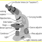
Diagram of a microscope The four main parts of a standard microscope are the light source, the objective lens, the stage, and the eyepiece.
The lens that is closest to the thing that has to be seen is the objective lens. Typically, this is a low-power lens with a 10x magnification. The platform that the specimen is set up on is called the stage. Since it is often movable, the user can reposition the specimen to get a more favorable angle.
The lens that allows you to view the specimen through is called an eyepiece. This lens often has a high power, about 40x magnification. The sample is illuminated by means of a light source. It is often situated beneath the stage and has adjustable brightness settings.
How a Microscope Operates
A microscope enlarges images through the use of lenses. The specimen is first amplified using the objective lens to create a picture, which is further enhanced by the eyepiece. The specimen is illuminated by a light source, which improves visibility.
The level of magnification that can be achieved with a microscope is limited by the quality of the lens and the amount of light that can be used. A typical microscope can achieve magnification of up to 1000x.
How to Use a Microscope
Using the microscope is relatively simple. First, place the sample on the stage and position it such that it is in focus. Next, adjust the light source so that it illuminates the sample evenly. Finally, look through the eyepiece and adjust focus until the image is clear.
How does the Light Source Work?
In a microscope, the light source is usually an LED (light-emitting diode). The LED shines a very bright light through the specimen and into the eyepiece. The light source is positioned such that the light passes through the objective lens before reaching the eyepiece.

How does the Objective Lens Work?
The objective lens is the lens closest to the object and is usually a high powered lens. The eyepiece is the lens farthest from the object. The light source shines through the specimen and is focused by the objective lens. The image is then magnified by the eyepiece.
How does the Eyepiece Lens Work?
Eyepiece lens in microscope diagram is one of the most important parts of the microscope. It is responsible for magnifying the image being viewed. The eyepiece lens is usually made of two lenses, which are placed in a metal tube. These lenses are known as objective lenses. The first lens, which is closest to the eye, is called the ocular lens. The second lens, which is farthest from the eye, is called the field lens.
The eyepiece lens works by bending and focusing the light rays passing through it. The eyepiece lens is able to do this because it has a curved surface. When light hits a curved surface, it bends. How much the light bends depends on how steep the curve is. The steeper the curve, the more the light is bent.
The eyepiece lens focuses the light by bringing all the light rays to a single point. This point is called the center point. The distance from the center of the eyepiece lens to the focal point is called the focal length. The shorter the focal length, the more powerful the microscope.
The eyepiece lens magnifies objects by making them appear closer than they actually are. It does this by increasing the apparent size of an object while maintaining its actual size relative to other objects. For example, if an object appears twice as large as another object when viewed through an eyepiece lens with a magnifying power.
Different Types of Microscopes
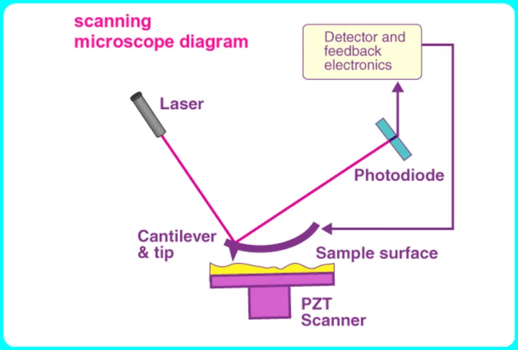
There are several varieties of microscopes, and each has a unique combination of characteristics and functionalities. The optical microscope, which magnifies objects using a lens, is the most popular kind of microscope. Other kinds of microscopes are the scanning probe microscope, which creates a three-dimensional picture by scanning an item with a sharp point, and the electron microscope, which magnifies an image using an electron beam.

FAQ
What is microscope class 8 biology?

With the aid of a microscope, one may examine tiny objects, including cells. The microscope has at least one lens that magnifies the picture of an item. By bending light in the direction of the eye, this lens inflates the size of an item.
Who is the father of microscope?

The founder of microscopy was Antoni van Leeuwenhoek (1632–1723).
What are the 5 uses of microscope?

Microscope Applications
They serve various functions in many sectors. Tissue analysis, forensic evidence evaluation, ecological health assessment, cellular function analysis, and atomic structure research are a few of their applications.
What is the function of microscope?

To see microorganisms that are too tiny to be seen with the human eye, a microscope is a necessary equipment. You must familiarize yourself with the primary portions of your microscope in order to operate it properly and efficiently in your everyday routine.
What are the 5 principles of microscope?

The fundamentals of microscopy, such as magnification, resolution, numerical aperture, lighting, and focusing, should be understood in order to use the microscope effectively and with the least amount of aggravation.
What is the structure of a microscope?
The primary components of a conventional biological microscope are the stage, reflector, lens tube, objective lens, and ocular lens. The objective lens enlarges an item that is presented on stage. Through the ocular lens, a magnified picture of the target may be seen after it is focussed.
What are the advantages of microscope?
Among a light microscope’s benefits are:
1. It enables us to view live things.
2. Staining methods are used in light microscopy to see various areas of a material.
3. A light microscope can magnify an object up to 1500 times its original size.


