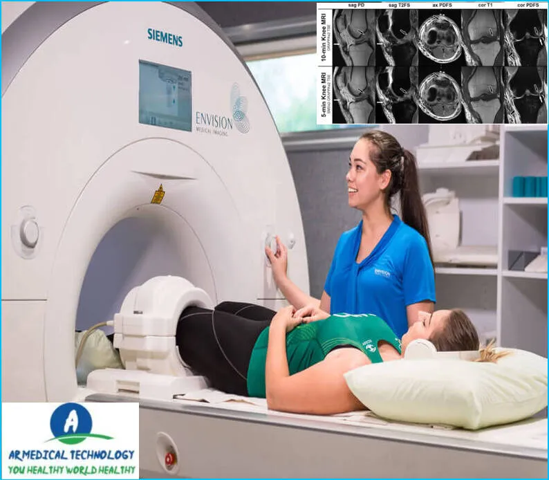
What to Expect During a Knee MRI
An MRI of the knee is a diagnostic technique that evaluates the health of the surrounding tissues and knee joint. MRI (magnetic resonance imaging) technology is used during this non-invasive treatment to get precise pictures of the knee. Numerous disorders, such as inflammatory arthritis, osteoarthritis, and rips in ligaments or tendons, can be diagnosed using a knee MRI.
On occasion, this process is also employed to assess the outcomes of prior knee surgery. You will lie on your back on an examination table with your leg extended for a knee MRI. You will be taken to an MRI scanner and have a wire wrapped around your leg. Throughout the exam, the equipment will produce a lot of noise, but you will always be able to speak with the technician over the intercom. Usually, the entire procedure takes thirty minutes or less. An MRI of the knee has no hazards, and there is no need to prepare for it beforehand.

How to Work Knee MRI
A magnetic resonance imaging (MRI) of the knee is a medical imaging method that shows the structures inside and surrounding the knee joint. It offers fine-grained pictures of ligaments, tendons, cartilage, and other soft tissues. This is a brief overview of what to anticipate and the usual procedure for a knee MRI:
Before the MRI:
- Preparation:
- Before the MRI, you may be asked to change into a hospital gown and remove any metal objects, including jewelry and accessories.
- Inform the technologist if you have any metal implants or devices in your body, such as pacemakers or metal joint replacements.
- Screening:
- You will undergo a screening process to ensure it’s safe for you to enter the magnetic field of the MRI machine.
- Patient Positioning:
- You will lie down on a moveable examination table, and the knee to be examined will be positioned in the center of the MRI machine.
During the MRI:
- Communication:
- You’ll be provided with earplugs or headphones to protect your ears from the loud noises produced by the MRI machine. Communication with the technologist is usually maintained through an intercom system.
- Immobilization:
- To obtain clear images, it’s important to remain as still as possible during the scan. Straps or foam pads may be used to help immobilize the knee.
- Contrast Agent (if used):
- In some cases, a contrast agent (dye) may be injected into a vein to enhance the visibility of certain structures. This is more common in cases where detailed visualization of blood vessels or soft tissues is required.
- Series of Scans:
- The MRI machine will produce a series of images, each lasting a few minutes. You may hear loud knocking or tapping sounds during the scan, which is normal.
After the MRI:
- Resume Normal Activities:
- In most cases, you can resume normal activities immediately after the MRI unless you were given a sedative.
- Results:
- The MRI images will be interpreted by a radiologist, and the results will be shared with your referring healthcare provider. They will discuss the findings and any necessary next steps with you.
Tips for a Successful Knee MRI:
- Follow any specific instructions provided by the healthcare team.
- Inform the technologist about any claustrophobia or anxiety you may have.
- Stay as still as possible during the scan to ensure clear images.
- If you have concerns or questions, don’t hesitate to communicate with the healthcare team.
Knee MRI
You may anticipate that the radiologist will obtain in-depth images of your knee during an MRI. They will utilize a device known as an MRI scanner to do this. These pictures are produced by radio waves and powerful magnets in an MRI scanner.

You will be required to complete a questionnaire on your medical history and current drugs prior to the surgery. Additionally, a consent form needs to be signed by you.
You will be required to remain still on a table during the process as the MRI machine captures images of your knee. In most cases, the process takes 30 to 60 minutes.
After the procedure, you will be able to go home and resume your normal activities. You should get a copy of the images from your MRI within a week.
There are no risks associated with an MRI of the knee.
Article About:- Health & fitness
Article About:- Medical Technology
Article About:- Sports
Knee MRI With or Without Contrast
It is possible to do a knee MRI with or without contrast. Contrast aids in improving the visibility of specific knee components. The process is the same whether contrast is used or not: On a table that glides into the MRI machine, you will lie on your back. An image-producing coil will be positioned around your leg. About 30 to 60 minutes are needed for the exam. An MRI of the knee, either with or without contrast, has no hazards.
Knee MRI Anatomy
A diagnostic technique for assessing the structure of the knee for anomalies is a knee MRI. The feet are tested by submerging them in a unique magnetic field-generating coil. This field generates fine-grained pictures of the knee in conjunction with radio waves.
You will lie on your back on a table with your legs in coils for a knee MRI. The table will be moved to the MRI machine’s center. In order to snap clear images during the exam, you will need to stay still.
torn meniscus normal knee mri vs abnormal
One frequent knee ailment is a torn meniscus. The meniscus is a piece of cartilage with a C shape that serves as a cushion between your knee’s bones. Meniscus tears may result in stiffness, edema, and discomfort.
Meniscal tears come in two varieties: degenerative and traumatic. Aging-related wear and tear is the cause of degenerative tears. Traumatic tears result from an injury, such a violent twist.
Conservative (non-surgical) methods, including as rest, ice, and physical therapy, are effective in treating the majority of meniscal injuries. Surgery could be necessary to heal certain rips, though.
You will lie on your back on a table with your leg straight out for a knee MRI. After that, the table will glide inside the MRI scanner. A magnetic field and radio waves are used in an MRI to produce finely detailed pictures of the inside of your body.
During the imaging process, you might be requested to hold your breath for a brief period of time in order to ensure quality photos.
You can go home right away after the surgery and get back to your regular routines. The use of MRI imaging has no dangers.
Knee MRI Cost
The price range for an MRI of the knee is $40 to $55. The cost will vary based on the MRI’s location, your insurance status, and if the procedure is covered by your plan. Should you be covering the cost out of pocket, you might be able to bargain with the imaging facility for a reduced fee.
An MRI of the knee is used to evaluate the health of the surrounding tissues and the knee joint itself. Meniscus injuries, stress fractures, arthritis, and rips in ligaments or tendons can all be diagnosed with the use of this imaging examination.

During an MRI of the knee, you will lie on your back on a table that slides into the center of the MRI machine. You will have to lie still during the procedure, which usually takes 30-60 minutes. There may be loud banging sounds during the scan, but you will be given earplugs or headphones to help block out these sounds.
After the process is complete, you will be able to go about your day as usual. You may experience some pain in your knee joint where the needle was inserted to inject the contrast material, but this should go away quickly.
There are no major risks associated with an MRI of the knee. However, people with metal implants or other metal objects in their body cannot have this type of scan due to safety concerns. Additionally, people with claustrophobia may find it difficult to remain still during the exam.
Knee MRI Procedure
An MRI of the knee is a diagnostic procedure that creates fine-grained pictures of the tissues inside and surrounding the knee joint using magnetic resonance imaging (MRI). This process is used to identify or rule out diseases such osteoarthritis, meniscus tears, and damaged ligaments.
You will lie on your back on an examination table with your leg extended for a knee MRI. Your thigh will have a cushioned block placed on top of it to stabilize your leg while the scan is being done. After positioning the scanner around your leg, the technician will start the scan. It’s common to hear loud banging noises when the scan is being performed. There may be headphones or earplugs available to assist block out sounds.
Knee MRI Risks
There are very few risks associated with knee MRI. The most common risk is that of allergic reaction to the contrast dye used in the procedure. There is also a small risk of infection at the injection site. Additionally, people with claustrophobia may experience anxiety while in the MRI machine.

FAQ
What does an MRI of a knee show?
Finding issues like damage to the ligaments and cartilage surrounding the knee might be aided by a knee MRI. Additionally, an MRI can be used to search for the source of infections in or around the knee, unexplained knee discomfort, and spontaneous knee failure.
How long does a knee MRI take?
Dye is occasionally injected into a joint. The radiologist can see certain locations more clearly because to the dye. The MRI operator will monitor you from a separate room during the procedure. Usually lasting between thirty and sixty minutes, the exam might take longer.
Is it OK to have an MRI with knee pain?
There are situations when transferred pain from another part of the body might cause knee discomfort. For instance, knee discomfort may be brought on by back or hip pain. In some situations, a knee MRI might not reveal any abnormalities, necessitating further testing to identify the underlying source of the discomfort.
Is MRI good for knee?
Which individuals with knee injuries need surgery can be identified with the use of MRI. When x-rays and other tests are not definitive, MRI can be used to assist detect a bone fracture. When using other imaging modalities, bone may hide anomalies that an MRI may identify.
Which is better for knee pain MRI or CT scan?
For the evaluation of soft tissue, chondral, and bony diseases of the knee joint, MRI is now the gold standard. SPECT/CT has been utilized and acknowledged for a growing number of orthopedic conditions within the past ten years. The primary use of SPECT/CT is the assessment of individuals who have persistent knee discomfort.
