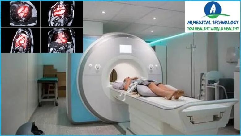
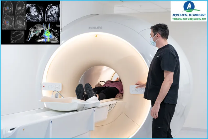
Cardiac Mri
A cardiac MRI, or magnetic resonance imaging, is a type of medical imaging that provides fine-grained pictures of the heart and its surrounding anatomy. It is an invaluable resource for the diagnosis and evaluation of a wide range of cardiovascular disorders since it offers thorough information about the composition and operation of the heart. An outline of a cardiac MRI’s uses and objectives is provided below:
1. Structural Assessment:
- Heart Chambers and Walls: Cardiac MRI provides high-resolution images that allow for a detailed examination of the heart’s chambers, walls, and valves.
- Pericardium: It can visualize the pericardium, the thin sac that surrounds the heart.
2. Functional Assessment:
- Cardiac Function: Cardiac MRI measures various parameters related to heart function, including ejection fraction (the percentage of blood pumped out with each heartbeat), stroke volume, and cardiac output.
- Valve Function: It assesses the functioning of heart valves, detecting abnormalities such as regurgitation or stenosis.
3. Blood Flow Dynamics:
- Blood Vessels: Cardiac MRI can visualize blood vessels and assess blood flow dynamics, helping identify abnormalities such as stenosis or aneurysms.
4. Tissue Characterization:
- Myocardial Tissue: Cardiac MRI provides information about the composition of myocardial tissue, aiding in the detection of abnormalities such as scar tissue, inflammation, or infiltration.
- Ischemic Heart Disease: It is useful for evaluating areas of reduced blood flow to the heart muscle, known as ischemia.
5. Assessment of Cardiac Masses:
- Tumors and Masses: Cardiac MRI can help identify and characterize cardiac masses, including tumors and other abnormalities.
6. Evaluation of Congenital Heart Disease:
- Congenital Anomalies: It is valuable in assessing congenital heart abnormalities and anomalies, providing detailed images for diagnosis and treatment planning.
7. Stress Cardiac MRI:
- Exercise or Pharmacological Stress Testing: Cardiac MRI can be used in stress testing scenarios to assess cardiac function and blood flow under conditions of increased stress.
8. Preoperative Planning:
- Cardiac Surgery: Cardiac MRI may be used for preoperative planning in cases where heart surgery is anticipated, providing detailed information for surgical guidance.
9. Monitoring Treatment Response:
- Follow-up Imaging: Cardiac MRI can be used to monitor the effectiveness of treatments and interventions, such as assessing changes in myocardial function after medical therapy or surgery.
Cardic MRI is a powerful diagnostic tool that offers detailed anatomical and functional information about the heart, making it particularly useful in cases where other imaging modalities may provide limited information. It is often employed in conjunction with other cardiovascular tests to provide a comprehensive evaluation of heart health. If recommended by a healthcare provider, a cardiac MRI is typically performed in a specialized imaging center by trained professionals.
Cardiac Mri
An MRI scan type called a cardiac MRI is used to evaluate the condition of the heart. An MRI machine is used to take images of the heart during the process. This enables the physician to assess the heart’s function and search for any anomalies.
Radiation is not used during the typically safe cardiac MRI technique. Nonetheless, there are certain dangers connected to it, like:
- Claustrophobia: Some people may feel anxious or claustrophobic when inside an MRI machine.
- Contrast dye: In some cases, a contrast dye may be used during the procedure. This may cause an allergic reaction in some people.
- Heart rate: The MRI machine can cause an increase in heart rate. This isn’t usually harmful, but it can be a problem for people who have heart disease.
Overall, cardiac MRI is a safe and effective method of assessing the health of the heart. This can help doctors diagnose problems sooner and start treatment sooner.
Article About:- Health & fitness
Article About:- Medical Technology
Article About:- Sports

Cardiac Mri With Contrast
A cardiac MRI with contrast is a type of MRI scan used to assess the heart and its blood vessels. Contrast agent helps to improve the quality of the images by making the structures of interest more visible. This procedure is generally safe, but there are some potential risks that should be considered before undergoing the test.
The purpose of a cardiec MRI with contrast is to evaluate the anatomy and function of the heart and its blood vessels. This information can be used to diagnose a variety of conditions such as coronary artery disease, cardiomyopathy, and valvular heart disease. In some cases, cardiac MRI may also be used to monitor the progress of these diseases or to assess the effectiveness of treatment.
The main risk associated with a cardic MRI with contrast is an allergic reaction to the contrast agent. This occurs in less than 1% of cases and is usually mild, resulting in a skin rash or hives. More severe reactions are rare, but can occur in people who already have allergies or who are particularly sensitive to the drugs. Another potential risk is nephrogenic systemic fibrosis, a rare condition that can develop in people with kidney problems after exposure to certain types of contrast agents.
If you’re considering having a cardic MRI with contrast, talk with your doctor about all the potential risks and benefits before scheduling the test.
Cost of Cardiac Mri
A cardiac MRI is a test that uses magnets and radio waves to create pictures of the heart. The procedure is also called magnetic resonance imaging (MRI) of the heart.
The cost of a Heart MRI can vary depending on a few factors, such as where you have the procedure done and whether you have insurance. According to NewChoiceHealth.com, the average cost of a cardiac MRI in the United States is around $2,000. However, if you do not have insurance, the cost can be as high as $5,000.
Cardiac Mri Cost
An MRI of the heart, also called a cardiec MRI, is a test that uses magnetic resonance imaging (MRI) to create pictures of the structures and blood flow in your heart. This type of MRI is different from other types because it uses a special machine and software to take pictures of your heart while it beats.
A cardiac MRI may be used to:
- look at the structure of your heart and valves
- assess your heart muscle function
- Find areas of damage or scarring in your heart muscle
- Find out how well treatments are working for conditions like coronary artery disease, cardiomyopathy, or valve disease
- Cardiac MRIs are usually performed as outpatient procedures. This means that you will not have to stay in hospital overnight. The process usually takes about an hour.
The cost of a cardiec MRI can vary depending on several factors, including who has the test done and whether you have insurance. Generally speaking, however, a cardiac MRI will cost between $1,500 and $3,000.
Cardiac Mri cpt Code
A cardiec MRI is a type of imaging test that uses magnetic resonance imaging (MRI) to create pictures of the heart. The test is also sometimes called cardiovascular magnetic resonance (CMR) or cardiac magnetic resonance imaging (CMRI).
The purpose of a cardiec MRI is to evaluate the structure and function of the heart. Cardiec MRI can be used to:
- Detect and identify coronary artery disease
- Evaluate heart muscle damage after a heart attack
- Assess the severity of valve problems
- detect abnormalities in the size or shape of the heart chambers
- evaluate for complications of congenital heart disease
- Plan a surgery to treat a heart condition
Cardiec MRI is generally safe and does not involve exposure to ionizing radiation. However, like all medical procedures, there are some risks associated with the test. These risks include:
Allergic reaction to contrast material
- kidney failure
- is bleeding
- arrhythmia
- abnormal heart rhythm
Rare but serious risks associated with the MRI procedure
Cardiac Mri Procedure
The Cardiec MRI is a diagnostic procedure that creates fine-grained pictures of the heart and its blood arteries using magnetic resonance imaging (MRI). The process is also occasionally referred to as cardiac magnetic resonance imaging, cardiac MR, or cardiovascular MRI.
You will recline on a table inside the MRI machine to have a cardiac MRI. For a little period, you will hold your breath while the equipment takes photos of your heart. 30 to 60 minutes are typically needed for the exam.
Cardiec MRI is used to assess the heart’s and its blood arteries’ anatomy and physiology. Cardiomyopathy (disease of the heart muscle) and coronary artery disease are among the ailments it can be used to identify. Cardiec MRI can also be used to evaluate how well certain disorders are being treated.
Heart magnetic resonance imaging carries extremely little risk. The process may cause you some discomfort because you will be lying motionless. Certain cardiac MRI treatments have a slight risk of having an allergic response to the contrast substance.

FAQ
What does a cardiac MRI diagnose?

Cardiovascular anatomical anomalies (like congenital heart defects), functional abnormalities (like valve failure), tumors, and conditions associated with coronary artery disease and cardiomyopathy (disease affecting the heart muscle) have all been successfully diagnosed using magnetic resonance imaging (MRI).
Can a cardiac MRI detect blockages?

Vivien Williams: An MRI may reveal to Dr. Shapiro various information, such as the location of blood clots, artery blockages, scar tissue, or even tumors, as well as damage from heart attacks or infections and the efficiency with which the heart pumps.
How long does a cardiac MRI take?

There are usually many employees in attendance. All of these individuals are engaged in cardiac MRI scanning. It takes 60 to 90 minutes for the scan. This is contingent upon both your heart rate and the test’s difficulty.
Is cardiac MRI better than CT scan?

Prof. Bamberg added, “MR is a good way to go when you need more information of the myocardium, but cardiac CT is sufficient for most patients to make good decisions.” Prof., CT is a perfect tool in a lot of situations.
What is the cost of a cardiac MRI?

The average cash cost for a cardiac MRI is $2,063, across all facilities. But depending on your location and any insurance coverage, the cost you pay varies a lot. To obtain the lowest prices and locate local providers of this service, enter your zip code.
Who needs a cardiac MRI?

When your doctor wants to rule out a heart condition, such as the source of your chest discomfort, shortness of breath, or fainting, they order a cardiac magnetic resonance imaging (MRI). a bigger heart. The cardiac muscle becomes thicker.
Is cardiac MRI better than echo?

Compared to echocardiography, cardiac magnetic resonance imaging (MRI) provides more information about soft tissues and can give specific insights on scarring, viability, and masses.
Which is better cardiac MRI or angiogram?

Cardiac MRI is recommended by doctors for a number of reasons, but it is particularly helpful when comprehensive pictures of the beating heart are required or as a noninvasive means of diagnosing heart muscle illness.
Is cardiac MRI expensive?

A cardiac MRI typically costs between $1,000 and $5,000, depending on a number of variables including the complexity of the ailment being looked into, the patient’s location, whether the procedure is being done in a hospital or a medical facility, and the kind of insurance the patient has.


